A comprehensive narrative review published in the Journal of the Egyptian National Cancer Institute synthesizes recent methodological advances and clinical translation challenges in applying explainable artificial intelligence to hepatocellular carcinoma research. Hepatocellular carcinoma represents a malignancy characterized by significant global incidence, mortality, substantial inter-tumor and intra-tumor heterogeneity at molecular, pathological, and imaging levels, and poor clinical prognosis. Although deep learning and machine learning models have demonstrated considerable diagnostic and prognostic accuracy across oncology workflows, their clinical deployment has been constrained by an opaque black-box architecture where decision-making logic remains concealed. The authors, Yuehua Li and Pengfei Li, examine how explainable artificial intelligence methods are transforming opaque predictions into interpretable findings across four principal clinical domains: imaging diagnosis, multi-omics biomarker discovery, treatment response prediction, and postoperative prognostic evaluation, while systematically outlining the pervasive technical bottlenecks that currently prevent post-hoc explanations from functioning as direct, actionable clinical interventions.
In radiological and digital imaging diagnosis, the primary function of explainable frameworks is visualizing model focus and aligning automated classifications with established clinical frameworks such as the Liver Imaging Reporting and Data System. Conventional deep learning architectures frequently produce unverified predictions; however, explainable techniques like Gradient-weighted Class Activation Mapping and SHapley Additive exPlanations decode internal feature activations into standardized clinical indicators, including non-rim arterial phase hyperenhancement. Beyond primary diagnostic categorization, explainable models non-invasively detect microscopic pathological phenomena directly from radiological scans. Representative investigations demonstrated that post-hoc attribution methods accurately highlighted the tumor margin and peritumoral boundary zones as decisive features for predicting microvascular invasion, aligning artificial intelligence outputs with histopathological realities. Furthermore, multi-task learning frameworks have linked routine macroscopic magnetic resonance imaging performances with underlying tumor grading, while specialized ultrasound and magnetic resonance sequence evaluations confirmed the adaptability of explainable systems across diverse imaging acquisitions, laying the technical groundwork for reliable multimodal diagnostic verification.
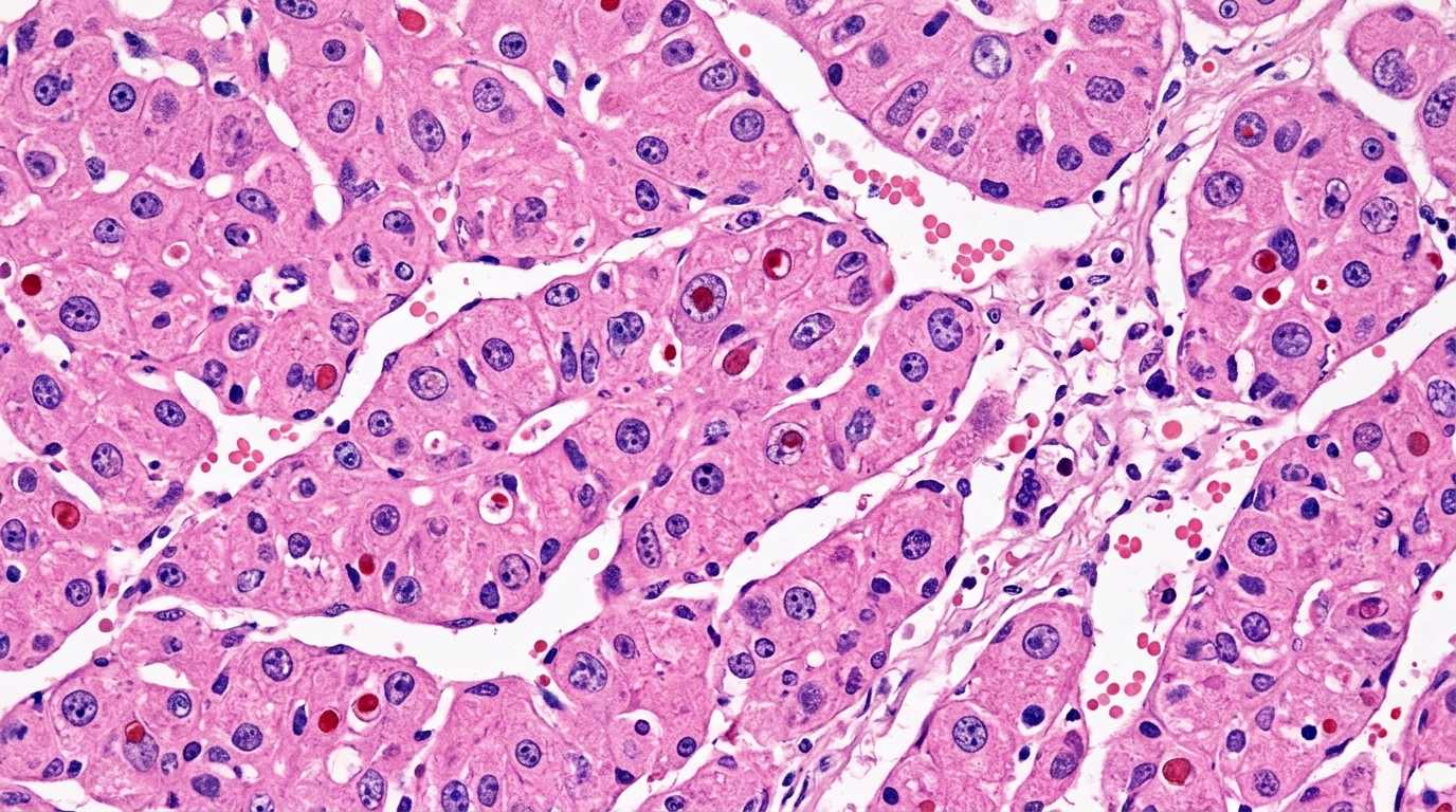
The scope of imaging-based explainability extends deeply into computational digital pathology and emerging spatial transcriptomic profiling. In whole-slide image analysis, explainable architectures apply attention-based visualizations and activation maps to render histological evaluations of nuclear atypia, tissue architectural distortion, and stromal alterations transparent, providing pathologists with an interpretable computational second opinion for tumor grading and subtype verification. This visual framework expands further as spatial transcriptomics generates high-resolution maps of molecular expression precisely registered to cellular tissue architecture. By linking macroscale imaging features to underlying cellular microenvironments, explainable systems have successfully decoded the biological foundations of specific magnetic resonance imaging phenotypes. This integration marks a critical evolution toward multi-scale explainability that unifies radiological, histopathological, and spatial molecular data layers into an auditable diagnostic continuum, directly addressing the multifaceted morphological heterogeneity characteristic of liver malignancies.
Within biomarker discovery and molecular subtyping, explainable artificial intelligence functions as a high-dimensional mining engine and hypothesis generator across complex multi-omics data layers, including genomics, single-cell transcriptomics, metabolomics, pathomics, radiomics, and routine clinical records. Rather than restricting analysis to isolated single-gene associations, explainable pipelines identify coordinated functional gene modules and biological pathways. Methodological frameworks utilizing tree-based feature attribution, automated machine learning, and multi-model feature selection have established reproducible pipelines to pinpoint robust candidate biomarkers while reducing false discoveries. Advanced implementations, such as intrinsically interpretable deep learning structures and pathway-directed post-hoc models, have successfully mapped prediction weights directly to known biological cascades, including the cyclic GMP-AMP synthase and stimulator of interferon genes signaling pathway, providing a mechanistic context that elevates computational predictions from ungrounded statistical patterns into testable biological hypotheses.
Pathomics and spatial molecular profiling further enrich this multi-omics discovery paradigm by converting routine histological tissue sections into quantitative phenotypic data. Interpretable deep learning models processing hematoxylin and eosin slides have successfully identified aggressive macrotrabecular structural patterns to predict microvascular invasion. Multimodal architectures such as token-guided fusion mechanisms and dual-branch exploratory networks simultaneously evaluate whole-slide morphological features, quantitative tumor-stroma ratios, and bulk gene expression profiles, explicitly calculating the individual predictive contribution of each respective biological layer. Moreover, quantification-based explainable techniques ground neural network activations directly within objective nuclear morphometric metrics, such as cellular size and pleomorphism, bridging computational discovery with standard pathology practice. In spatially resolved transcriptomics, explainable algorithms pinpoint microenvironment signatures within specific histological regions, clarifying the spatial determinants of therapeutic resistance and immune cell infiltration patterns across distinct tumor niches.
In the evaluation of therapeutic efficacy, explainable models provide critical insights into treatment response heterogeneity across locoregional and systemic interventions, facilitating a shift away from standardized protocols toward individualized care. In transarterial chemoembolization, attribution modeling with SHapley Additive exPlanations has unraveled the underlying drivers of variable patient responses. Studies analyzing pre-treatment contrast-enhanced computed tomography parameters and dynamic trajectory tracking of albumin-bilirubin scores have identified distinct responder subtypes, highlighted patients unsuited for primary transarterial chemoembolization, and revealed that splenic and renal imaging characteristics significantly inform outcome estimates by reflecting systemic inflammatory states. Similarly, for systemic therapies encompassing targeted agents and immune checkpoint inhibitors, explainable models have linked baseline radiographic features and cellular pathomic signatures, such as tumor-infiltrating lymphocyte density, to therapeutic responsiveness and specific molecular pathways, while other models have mapped hepatic toxicity risks following radiotherapy and ablation to balance treatment efficacy against procedural safety.
For postoperative prognosis and long-term surveillance, explainable frameworks have achieved refined risk stratification by integrating standard clinical metrics with advanced radiomics and multi-omics indicators. Multimodal nomograms and random survival forest models incorporate computed tomography and magnetic resonance radiomics to quantify the biological invasiveness of tumors, reliably forecasting postoperative recurrence in hepatitis B-related malignancies and large hepatocellular carcinomas. Explainable models tailored to specific sub-populations have demonstrated equal clinical precision, providing personalized outcome forecasts for alpha-fetoprotein-negative patients, individuals with concurrent diabetes mellitus, and patients at elevated risk for spontaneous tumor rupture or post-hepatectomy liver failure. Incorporating temporal longitudinal features, such as fluctuating liver function trajectories and post-cure hepatitis C viral monitoring, aligns artificial intelligence capabilities with the ongoing clinical requirements of chronic liver disease management.
Despite these extensive advancements, the review highlights three fundamental methodological bottlenecks inherent to the prevailing post-hoc explanation paradigm that severely constrain its translational utility. First, post-hoc methods remain fundamentally correlational rather than causal; they highlight statistical dependencies within historical datasets without confirming whether highlighted features represent genuine biological drivers or incidental epiphenomena. Second, a persistent semantic gap separates low-level, pixel-oriented saliency heatmaps from the high-level semantic reasoning utilized by clinicians, leaving models vulnerable to shortcut learning where predictions rely on artifacts rather than disease mechanisms. Third, current predictive architectures operate as static snapshots trained on historical cohorts, rendering them incapable of self-updating alongside evolving clinical guidelines or actively comparing alternative therapeutic outcomes.
To overcome these limitations, the authors delineate a paradigm shift centered on next-generation technical architectures: moving toward causal inference, concept-driven interpretability, and dynamic collaborative systems. Methodologies such as double machine learning and causal forests provide mathematical rigor to control for high-dimensional confounding, enabling the estimation of true treatment effects and identifying heterogeneous patient cohorts that derive differential survival benefits from specific interventions. Concept Bottleneck Models offer a viable architectural solution to bridge the semantic divide by forcing intermediate network layers to predict human-interpretable clinical concepts, such as arterial phase washout or histological nuclear atypia, prior to generating final classifications. Furthermore, mechanistic interpretability techniques utilizing sparse autoencoders show promising capability in disentangling the complex, polysemantic latent representations of large-scale pathology foundation models into individual, monosemantic biological concepts.
The review concludes by emphasizing that the ultimate maturation of explainable artificial intelligence depends on transitioning from passive, retrospective explanatory tools into active, interactive clinical decision partners. Incorporating continual learning algorithms will allow diagnostic and prognostic models to integrate real-time patient trajectories and updated institutional regimens without succumbing to catastrophic forgetting of previously acquired knowledge. Simultaneously, coupling counterfactual explanation generators with case-based retrieval systems will allow practitioners to interactively explore alternative clinical scenarios, answering critical questions regarding how potential therapeutic interventions might alter an individual patient’s prognosis. Realizing this clinical transition will require overcoming pragmatic accessibility hurdles, including resource shortages in digital pathology infrastructure, elevated deployment expenditures, and workflow integration hurdles, alongside rigorous prospective clinical validation to ensure that transparent computational models truly deliver safer, personalized precision oncology.
Subject of Research: Explainable artificial intelligence applications, limitations, and future paradigms in hepatocellular carcinoma research
Article Title: From black-box prediction to transparent insight: the status quo and paradigm shift of explainable artificial intelligence in hepatocellular carcinoma research
Article References: Li, Y., & Li, P. (2026). From black-box prediction to transparent insight: the status quo and paradigm shift of explainable artificial intelligence in hepatocellular carcinoma research. Journal of the Egyptian National Cancer Institute, 38(1), Article 59. https://doi.org/10.1186/s43046-026-00397-0
Image Credits: AI Generated
DOI: 10.1186/s43046-026-00397-0
Keywords: Hepatocellular Carcinoma, Explainable Artificial Intelligence, Deep Learning, LI-RADS, Grad-CAM, SHAP, Concept Bottleneck Models, Digital Pathology, Multi-Omics, Microvascular Invasion, Transarterial Chemoembolization, Radiomics, Double Machine Learning, Causal Forests, Continual Learning, Counterfactual Explanations, Spatial Transcriptomics
News Source: Nathaniel Bowman. (November 20, 2026). Explainable Artificial Intelligence Advances Hepatocellular Carcinoma Research Toward Actionable Clinical Insight. Scienmag.