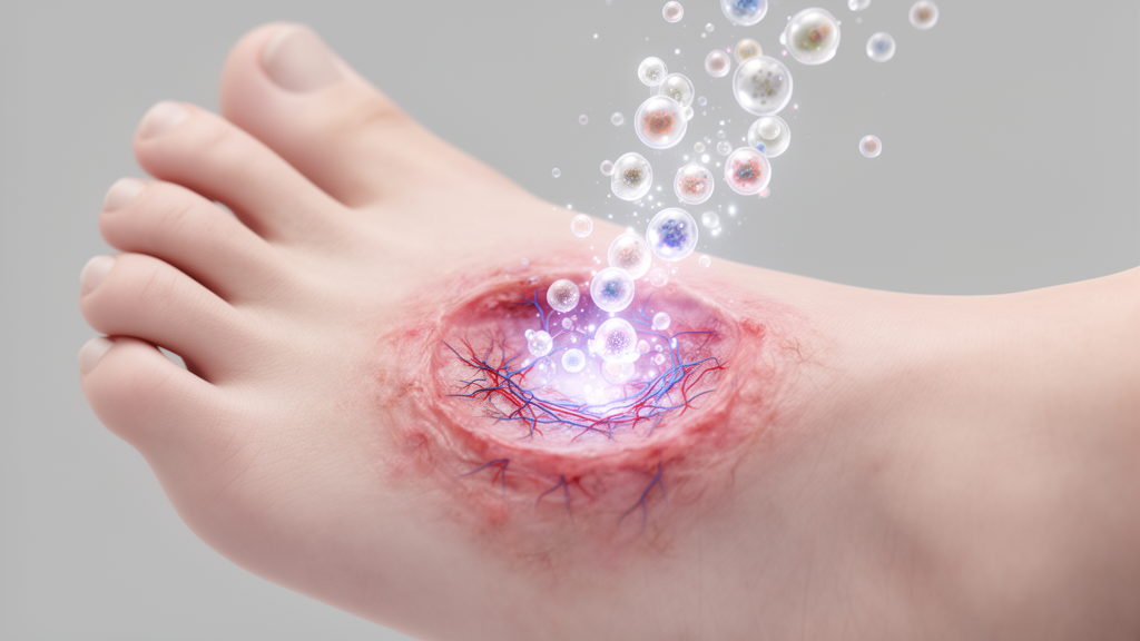
Diabetic foot ulcers remain one of the most stubborn and clinically consequential complications of diabetes, affecting millions of patients worldwide and often resisting every conventional treatment from advanced dressings to surgical debridement. A newly published review in the journal Skin takes a hard look at one of the most talked-about emerging therapies in regenerative medicine: extracellular vesicles, commonly known as exosomes or small extracellular vesicles (sEVs). The review, titled “Exosome-Mediated Regulation of Re-Epithelialization and Epithelial–Mesenchymal Plasticity in Diabetic Cutaneous Wounds: Current Advances and Translational Perspectives,” was published on September 3, 2026, and delivers a message that is both encouraging and sobering. These nanoscale vesicles, shed by nearly every cell type in the body, appear capable of improving multiple aspects of the diabetic wound environment. Yet when it comes to the single most important question—whether they directly orchestrate the cellular program that rebuilds the skin’s protective barrier—the evidence, the authors conclude, is still strikingly thin.
To understand why this gap matters, it helps to appreciate what actually happens when a wound closes. The outermost layer of the skin, the epidermis, is restored through a process called re-epithelialization, which depends on keratinocytes, the dominant cell type of the epidermis, located at the wound edge. These cells must undergo a remarkable sequence of behaviors: they activate, detach from their neighbors, migrate across the wound bed, proliferate to replenish the migrating front, and then—critically—redifferentiate back into their mature state, reassembling cell junctions and restoring the layered barrier structures that keep microbes out and moisture in. Each stage of this choreography is vulnerable to disruption, and in diabetes, the high-glucose environment and its downstream pathological consequences interfere at nearly every step, which is why diabetic wounds so often stall in a chronic, non-healing state.
One of the review’s most important conceptual contributions is its argument about how this process should be described in the first place. For years, wound biologists have leaned on the framework of epithelial–mesenchymal transition, or EMT, a binary model in which epithelial cells are thought to convert wholesale into migratory, mesenchymal-like cells and then switch back. The authors contend that this binary picture does not accurately capture what keratinocytes actually do during repair. Instead, they argue, the phenomenon is better described as epithelial–mesenchymal plasticity, or EMP—a spectrum of transient, reversible, and stage-dependent phenotypic states rather than an all-or-nothing switch. This distinction is not merely semantic. If keratinocytes occupy a continuum of hybrid states during migration and then slide back toward full epithelial identity as the barrier reforms, then therapies must be evaluated not just for whether they speed up closure, but for whether they support the full, coordinated sequence of state changes, including the final redifferentiation and junction reassembly that produce a functional barrier.
Exosomes and other small extracellular vesicles have captured the imagination of wound-healing researchers precisely because they are nature’s own intercellular delivery vehicles. These lipid-enclosed nanoparticles carry proteins, lipids, and RNA molecules from their cell of origin to recipient cells, and in doing so they can reprogram the behavior of the cells they reach. In the context of diabetic wounds, the review explains, sEVs may support re-epithelialization through two broad routes. The first is direct: vesicles could act on keratinocytes themselves, influencing their repair programs—promoting migration, stimulating proliferation, and potentially guiding the redifferentiation and junction formation needed for barrier recovery. The second is indirect: vesicles derived from stem cells, immune cells, or other sources could remodel the hostile wound microenvironment, dampening chronic inflammation, encouraging new blood vessel growth, reducing oxidative stress, and improving the extracellular matrix scaffold on which migrating keratinocytes depend.
The indirect route is where the bulk of the experimental evidence currently sits, and this is the crux of the review’s critique. Most published studies of exosome or sEV therapy in diabetic wound models measure endpoints such as overall wound closure, histological re-epithelialization, angiogenesis, inflammatory marker changes, or collagen deposition. These outcomes are meaningful, and improvements in them justify continued enthusiasm for the approach. But they are, by their nature, downstream or parallel consequences of therapy. They do not tell researchers whether the vesicles actually engaged the keratinocyte plasticity program itself—whether they nudged cells through the EMP-associated state transitions, supported epithelial redifferentiation, and produced genuine, durable barrier recovery in diabetic wound keratinocytes. On that direct question, the authors find the evidence remains limited, and they are candid about the implications for how the field should interpret its own successes.
To bring analytical clarity to a literature of uneven depth, the review proposes a four-tier framework for grading the evidence. The highest tier is direct EMP evidence: studies that track the actual phenotypic state transitions of keratinocytes in diabetic wounds under sEV treatment. The second tier is functional keratinocyte evidence: demonstrations that vesicles measurably alter keratinocyte migration, proliferation, redifferentiation, or barrier-forming capacity, even if the full plasticity trajectory is not mapped. The third tier is indirect microenvironmental support: the well-populated category of studies showing improved inflammation, vascularization, oxidative balance, or matrix remodeling. The lowest tier is mechanistic inference, where benefits are observed but the cellular mechanism is assumed rather than demonstrated. This hierarchy gives researchers, reviewers, and clinicians a shared vocabulary for assessing how mature any given line of evidence really is—a tool the field has arguably lacked as publications on vesicle therapies have multiplied.
The review also confronts the practical and translational dimensions that will determine whether laboratory promise becomes bedside reality. Engineered vesicles—vesicles whose cargo or surface properties have been deliberately modified to enhance potency, targeting, or stability—represent one active frontier, offering the possibility of tailoring sEV therapies to specific stages of the repair sequence. Local delivery systems are another: because free vesicles can diffuse away, degrade, or be cleared rapidly from a wound bed, biomaterial-based depots, hydrogels, and scaffolds designed to retain and release vesicles at the wound site are being explored as ways to sustain therapeutic concentrations. Yet each of these strategies introduces its own questions about manufacturing consistency, dose standardization, safety, and regulatory classification, and the review treats evidence maturity and translational risks as inseparable from the biology itself.
The stakes of getting this right are considerable. A therapy that accelerates wound closure but leaves behind a fragile, poorly differentiated epidermis could set patients up for re-ulceration, the dreaded cycle that defines much of diabetic foot disease. Conversely, a therapy that genuinely guides keratinocytes through the full EMP arc—from activation and migration through redifferentiation and junction restoration—would address the fundamental cellular failure of chronic diabetic wounds rather than merely palliating the environment around it. This is why the authors insist that future studies move beyond wound-area measurements and incorporate readouts of keratinocyte state, epithelial stratification, and barrier function. The distinction between a closed wound and a healed one is, in diabetic skin, the difference between temporary reprieve and durable recovery.
For now, the review’s overall verdict is a calibrated one. Exosome and small extracellular vesicle therapies have earned their place among the most promising strategies for diabetic wound healing, with plausible mechanisms spanning direct keratinocyte support and broad microenvironmental remodeling. The multiple microenvironmental effects documented across preclinical studies are real and encouraging. But the field’s central claim—that these vesicles can deliberately guide the plasticity program that rebuilds the epidermal barrier—still rests more on inference than on direct demonstration. By naming the gap precisely, proposing a framework to close it, and mapping the engineering and delivery challenges ahead, the review offers the field something more valuable than another positive result: a clear picture of what must be proven before exosome therapy can be trusted to heal, and not merely close, the wounds of diabetic patients.
As vesicle-based therapeutics advance toward clinical testing, the EMP framework articulated in this review is likely to shape how the next generation of experiments is designed. Studies that track keratinocyte state transitions directly, validate functional barrier recovery, and report evidence maturity transparently will be the ones that determine whether exosome therapy fulfills its promise for the patients who need it most—those whose wounds have run out of options.
Subject of Research: Exosome and small extracellular vesicle therapy for diabetic wound re-epithelialization and epithelial–mesenchymal plasticity
Article Title: Exosome/sEV therapy for diabetic wounds: Promise and evidence gaps in re-epithelialization
Article References: Exosome/sEV therapy for diabetic wounds: Promise and evidence gaps in re-epithelialization. Original publication (Bibliographic details unavailable.)
Image Credits: AI Generated
DOI: Not provided
Keywords: exosomes, small extracellular vesicles, diabetic foot ulcers, wound healing, re-epithelialization, keratinocytes, epithelial-mesenchymal plasticity, epidermal barrier, regenerative medicine, stem cell therapy, drug delivery, Skin journal
News Source: Drew Townsend. (October 19, 2026). Exosome Therapy Shows Promise for Diabetic Wounds, but Key Evidence Remains Scarce. Scienmag.