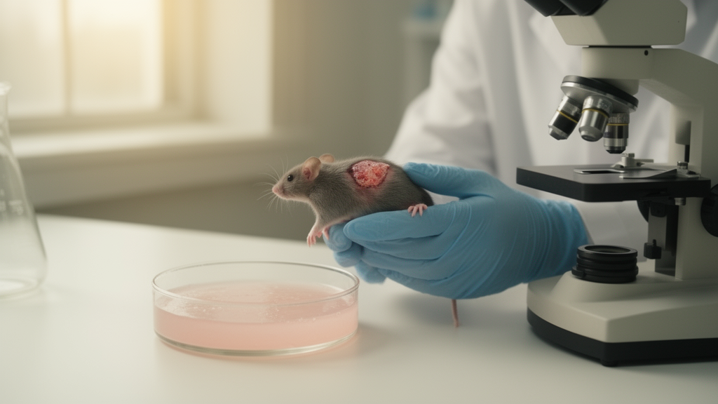
Researchers have engineered a recombinant human type I collagen that keeps the stable triple-helical shape of natural collagen while being produced entirely in bacteria, and they report that a topical version of the protein accelerated recovery of sun-damaged skin in mice. The material, called triple-helical recombinant humanized type I collagen, or THRCI, was described in a study published on 1 June 2026 in the journal Collagen and Leather. In a mouse model of acute ultraviolet photodamage, daily topical application of the protein sped up epidermal recovery, restored skin hydration and barrier function, raised dermal density back toward baseline, and promoted remodeling of collagen fibers in the dermis. At the same time, the treatment lowered expression of the inflammatory signals IL-6 and IL-1β and of the tissue-degrading enzymes MMP-1 and MMP-9 in the damaged skin.
The motivation behind the work lies in a long-standing gap between what collagen can do in skin repair and what most available collagen products can actually deliver. Collagen is the main structural protein of the dermis, and its mechanical stability comes from a repeating Gly-X-Y amino acid motif that allows three collagen chains to wind together into a tight triple helix. That conformation gives the protein tensile strength and also presents functional motifs, such as the sequence GFPGER, that bind to integrin receptors on cells and regulate adhesion, migration, and differentiation. Type I collagen, the predominant form in skin, is a cornerstone biomaterial in tissue engineering. Yet collagen extracted from animal sources such as tendon or skin can carry risks of immunogenicity and pathogen transmission, and extraction itself often compromises the triple-helical integrity that underlies the protein’s bioactivity.
Recombinant collagen, made through genetic engineering, avoids batch variability, ethical concerns, and potential viral contamination, and it has been produced in mammalian cells, yeast, transgenic plants, and Escherichia coli. The bacterium remains attractive because of its high yield, ease of genetic manipulation, and low cost. But recombinant type I collagen made in E. coli has historically fallen short in two ways: the triple-helix structure is incomplete, and thermal stability is poor. Systems that co-express the enzyme prolyl-4-hydroxylase with collagen chains in E. coli produce partially hydroxylated collagens with better helical stability, but those systems are hard to control precisely, burden the host cell’s metabolism, and complicate scale-up. The team behind THRCI took a different route, designing the protein sequence itself so that the helix is inherently stable without depending on enzymatic modification during production.
The amino acid sequence of THRCI is derived from the human type I collagen α1 chain and preserves the canonical Gly-X-Y repeats and the GFPGER motif needed for receptor binding. The designed protein contains an N-terminal globular V-domain fused to the collagenous triple-helical domain, with a protease cleavage site between them so the V-domain can be removed after purification. The gene was synthesized and inserted into the pCold III vector under a cold-shock promoter, then expressed in E. coli BL21(DE3). Fermentation was scaled to a 100-liter bioreactor, and the researchers used a temperature strategy to improve folding: after induction at an optical density of 30.0, the culture was held at 25 degrees Celsius for 12 hours and then lowered to 20 degrees Celsius for another 12 hours. This approach yielded approximately 2.0 grams of THRCI per liter of culture.
Purification relied on a histidine tag and nickel-affinity chromatography, followed by thrombin digestion to cleave off the V-domain and dialysis to remove the fragments. Characterization confirmed that the final protein behaved like native collagen. Mass spectrometry measured the molecular mass at 15,104.0 daltons, closely matching the theoretical value of 15,099.7 daltons. Circular dichroism spectroscopy showed the characteristic positive peak near 220 nanometers and negative peak around 198 nanometers that signal a triple helix, and thermal unfolding monitored at 220 nanometers gave a melting temperature of 37 degrees Celsius, essentially body temperature. Fourier transform infrared spectroscopy found amide I, II, and III absorption bands at 1655, 1554, and 1240 wavenumbers, consistent with type I collagen, and heat denaturation erased the characteristic amide I signature, replacing it with a single broad band. A protease sensitivity test provided perhaps the most direct evidence of helical integrity: native THRCI resisted trypsin digestion for 24 hours, retaining 84.4 percent of its band intensity, while heat-denatured THRCI was completely degraded.
In laboratory tests with human foreskin fibroblasts, THRCI supported the cellular behaviors that matter for tissue repair. Cell viability stayed above 100 percent across concentrations from 0.001 to 1.0 milligrams per milliliter, indicating no cytotoxicity. Over five days, proliferation of THRCI-treated fibroblasts rose to 122.19 percent of the blank control, statistically indistinguishable from cells grown on native collagen extracted from yak tendon. Cells cultured on THRCI-coated surfaces adopted the spindle-shaped morphology characteristic of adherent fibroblasts, unlike the spherical cells seen on a bovine serum albumin negative control, an effect the authors attribute to the GFPGER integrin-binding motif. In scratch assays, wounds closed at a rate of 51.35 percent after 24 hours in the THRCI group versus 19.04 percent in untreated controls, again matching the native collagen comparison. After seven days of co-culture, THRCI significantly increased expression of α-SMA, a marker of fibroblast-to-myofibroblast differentiation, along with type I and type III collagen genes, indicating enhanced extracellular matrix synthesis.
The researchers then tested whether THRCI could protect cells from ultraviolet-induced oxidative damage. Because UVB primarily affects keratinocytes while UVA predominantly targets fibroblasts, they used UVB-irradiated HaCat keratinocytes and UVA-irradiated HFF-1 fibroblasts. Ultraviolet exposure drove a marked rise in intracellular reactive oxygen species, detected by fluorescent staining and confirmed by flow cytometry, but two hours of pretreatment with THRCI significantly reduced ROS levels in both cell types. The treatment also raised activity of the antioxidant enzyme superoxide dismutase, to 2.37-fold of the damaged control level in keratinocytes and 1.54-fold in fibroblasts, and it suppressed accumulation of malondialdehyde, a terminal byproduct of lipid peroxidation. In zebrafish larvae exposed to copper sulfate to induce oxidative stress, THRCI reduced ROS fluorescence to 33.58 percent of the stressed control, comparable to the established positive control dipotassium glycyrrhizinate at 35.17 percent.
Zebrafish also provided a living model for the anti-wrinkle question. Larvae were incubated with THRCI and then given three cycles of UVB irradiation, and the area of the caudal fin was quantified 22 hours later. Ultraviolet exposure shrank the fin area to 55.16 percent of the unirradiated blank control, but larvae treated with THRCI retained 87.77 percent of their fin area, essentially matching both the native collagen group at 87.72 percent and the tea polyphenol positive control at 92.76 percent. Safety testing in rabbits found no skin irritation from THRCI dressings, with a primary irritation index of zero; a hemolysis rate of 0.98 percent, well under the 5 percent threshold for blood-contacting biomaterials; and a cumulative temperature rise of 0.9 degrees Celsius after intravenous administration, within the pyrogenicity limits of the Chinese Pharmacopoeia.
The central experiment used Kunming mice whose shaved dorsal skin was exposed to UVA and UVB radiation totaling 200 joules per square centimeter, followed by daily topical application of 5 milliliters of a 0.5 milligram per milliliter THRCI solution. Measurements with a DermaLab Combo system tracked recovery over seven days. On day one, dermal density in the UV-damaged groups had collapsed from a blank-group value of 62.01 to between 21.41 and 28.46. By day seven, dermal density in the THRCI group had climbed to 59.72, statistically indistinguishable from the blank group at 63.41, while the saline-treated model group remained at 37.35. Hydration, which fell from 265.65 in blank animals to roughly 114 to 125 in all damaged groups on day one, was fully restored in the THRCI group by day seven, as was transepidermal water loss, a marker of barrier integrity. Videoscope imaging showed scabs detaching by day five and erythema fully resolved by day seven in the treated groups, while the model group still showed notable redness.
Histology and gene expression analysis filled in the mechanism. Hematoxylin and eosin staining showed that by day seven, epidermal thickness in the THRCI group was statistically indistinguishable from uninjured skin, with intact dermal-epidermal junctions, whereas the model group retained persistent epidermal hyperplasia and blurred boundaries. Masson staining quantified collagen volume fraction: after UV damage it dropped to around 28 to 34 percent across damaged groups, but by day seven the THRCI group reached 45.08 percent and the native collagen group 46.22 percent, both comparable to blank skin, while the model group lagged at 31.75 percent. At the transcript level, THRCI reduced IL-6 to about 0.74-fold of the model level and IL-1β to roughly 0.73-fold, cut MMP-1 to approximately 0.51-fold and MMP-9 to about 0.76-fold, and raised Collagen I expression 1.53-fold and Collagen III 1.64-fold relative to the damaged control. The authors conclude that THRCI accelerates skin regeneration by reducing inflammation, suppressing matrix-degrading enzyme expression, and enhancing collagen synthesis. Because all efficacy data come from cell cultures, zebrafish larvae, and a mouse model rather than human trials, the findings establish preclinical potential, and the researchers position THRCI as a candidate for clinical translation in skin repair and rejuvenation therapies.
Subject of Research: Development and preclinical evaluation of a triple-helical recombinant humanized type I collagen for repairing ultraviolet-damaged skin
Article Title: Triple-helical recombinant humanized type I collagen promotes photodamaged skin repair via ECM remodeling and anti-inflammatory mechanisms
Article References: Wei, N., Zhang, Y., Huang, W., Tian, X., Yao, L., & Xiao, J. (2026). Triple-helical recombinant humanized type I collagen promotes photodamaged skin repair via ECM remodeling and anti-inflammatory mechanisms. Collagen and Leather, 8(1), Article 23. https://doi.org/10.1186/s42825-026-00245-w
Image Credits: AI Generated
DOI: 10.1186/s42825-026-00245-w
Keywords: recombinant collagen, triple helix, type I collagen, photoaging, UV photodamage, skin repair, extracellular matrix, anti-inflammatory, E. coli expression, fibroblasts, zebrafish model, biomaterials
News Source: Denise Maddox. (October 30, 2026). Engineered human collagen with a stable triple helix speeds skin repair in mice after UV damage. Scienmag.