In a groundbreaking study that illuminates the delicate balance between speed and precision in early embryonic development, researchers at Hokkaido University have unveiled critical insights into how zebrafish embryos manage the complex process of cell division under stress. Led by Professor Ryota Uehara, the team employed cutting-edge optochemical techniques to selectively disrupt mitosis—the process by which cells divide—at distinct developmental stages. Their findings, published in Communications Biology on March 23, 2026, reveal a previously unappreciated tolerance to cellular errors during the gastrula stage of development, advancing our understanding of embryogenesis and opening novel avenues for cancer research.
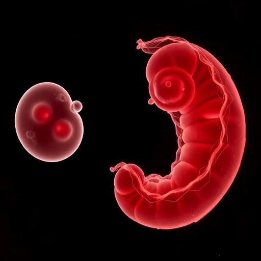
The early development of an embryo demands rapid cell division to enable swift growth, yet this speed comes with an inherent risk: the propagation of errors during mitosis can be catastrophic, potentially derailing subsequent developmental processes. Embryonic cells must therefore navigate this precarious trade-off, balancing the urgency of proliferation with the necessity of genomic integrity. Professor Uehara’s research focuses on this tightrope walk, utilizing zebrafish embryos as a model system due to their transparent nature and genetic similarity to other vertebrates.
Central to the process of mitosis is the alignment and segregation of chromosomes, orchestrated by a sophisticated cellular machinery. Chromosomes, paired and duplicated, need to be precisely lined up at the metaphase plate and subsequently pulled apart by spindle fibers to ensure each daughter cell inherits an exact copy of genetic material. Disruption in this choreography is often lethal, as it can result in aneuploidy or other chromosomal abnormalities. The protein centromere-associated protein E (CENP-E) plays a pivotal role here by facilitating the accurate alignment of chromosomes. The research team harnessed a light-activated chemical inhibitor to transiently suppress CENP-E activity, introducing deliberate errors to probe the resilience of embryonic developmental stages.
The use of optochemical methods represents a significant technological advance in developmental biology. By applying precisely controlled pulses of light, researchers can regulate biological processes with exquisite spatial and temporal resolution. This allowed the investigators to target CENP-E function at specific developmental windows and examine the resultant effects on embryogenesis. The approach also circumvents the systemic toxicity and lack of temporal specificity associated with traditional genetic knockouts or pharmaceutical inhibitors.
Strikingly, the study delineated a critical window just prior to gastrulation—a phase when the embryo transitions from a single-cell-thick layer to a multilayered structure—during which embryos are particularly vulnerable to mitotic disruption. When CENP-E activity was inhibited during this pre-gastrula phase, the embryos exhibited high mortality rates, especially when the inhibition was sustained over multiple cell cycles. This finding underscores the paramount importance of flawless chromosome alignment when cellular architecture is relatively simple and undifferentiated.
Conversely, embryos that had progressed to the gastrula stage demonstrated a remarkable capacity to endure prolonged CENP-E inhibition. This robustness suggests an adaptive mechanism that mitigates the consequences of mitotic errors despite ongoing chromosomal misalignment. The research team unveiled that this tolerance hinges on the spindle assembly checkpoint (SAC), an intrinsic surveillance system that monitors kinetochore-microtubule attachments and delays anaphase onset until chromosomes achieve at least partial alignment. The SAC in gastrula-stage embryos, while less stringent than that in adult cells, appears sufficiently robust to prevent fatal errors during division.
To further probe this survival mechanism, researchers introduced simultaneous inhibition of both CENP-E and the spindle assembly checkpoint. This dual disruption proved devastating, causing catastrophic embryonic lethality even at the gastrula stage. The synergy of impairing chromosome alignment and the checkpoint’s monitoring ability underscores the SAC’s crucial role as a cellular safeguard during periods of high developmental complexity and stress.
Beyond its fundamental contribution to developmental biology, this research holds profound implications for oncology. CENP-E has long been considered a promising target for anti-cancer therapeutics due to its essential function in mitosis. The ability to modulate its activity with light-sensitive molecules opens new horizons for precisely targeted cancer therapies that minimize collateral damage to healthy tissues. Professor Uehara envisions future clinical applications where optochemically controlled inhibitors could selectively suppress tumor cell proliferation with unprecedented accuracy, harnessing spatial and temporal light control.
The researchers’ success in demonstrating optochemical control over CENP-E in a live vertebrate embryo marks a technological and conceptual milestone. It validates the potential of integrating chemical biology with photonics to dissect and manipulate complex biological systems in vivo. This approach furnishes a powerful platform for exploring dynamic processes in normal development and disease models, enabling interventions that were previously impossible with conventional methods.
The study’s findings also challenge existing paradigms concerning the embryonic cell cycle and its regulatory checkpoints. Traditionally, early embryos were thought to operate under a more permissive cell cycle regime, prioritizing speed over error correction. However, the identification of an operational spindle assembly checkpoint during the gastrula phase suggests a nuanced, stage-dependent modulation of mitotic fidelity. This adaptability is likely essential for balancing the competing demands of rapid growth and genomic accuracy.
Comprehensive genetic and biochemical analyses are anticipated to follow, aimed at elucidating the molecular underpinnings of this incomplete checkpoint. Understanding how the SAC modulates its stringency and interacts with other cell cycle regulators could unlock new strategies for manipulating cell division in both developmental and pathological contexts. In particular, insights gleaned from zebrafish models may translate into innovative approaches for cancer treatment, regenerative medicine, and tissue engineering.
Professor Uehara’s team continues to refine their optochemical toolset to achieve higher precision and versatility, exploring other mitotic proteins and checkpoints. As researchers expand the optogenetic and chemical biology toolbox, these advancements promise to transform not only developmental biology but also broader biomedical fields. The pioneering work outlined herein is a vivid testament to the power of interdisciplinary science in unraveling life’s complexity, offering hope for improved therapeutic interventions against diseases marked by aberrant cell division.
In sum, the elucidation of the spindle assembly checkpoint’s role in zebrafish embryonic development via photochemically controlled CENP-E inhibition represents a paradigm shift in our understanding of early developmental biology. This study highlights the embryo’s evolving capacity to cope with division errors and provides a foundation for future innovation in both basic science and clinical cancer therapies. The fusion of optical control and chemical biology heralds a new era of dynamic, precise manipulation of biological systems with vast implications for human health.
Subject of Research: Cells
Article Title: Optochemical elucidation of a critical role of the incomplete spindle assembly checkpoint in zebrafish development
News Publication Date: 23-Mar-2026
Web References:
http://dx.doi.org/10.1038/s42003-026-09871-w
Image Credits: Uehara Lab, Faculty of Advanced Life Science, Hokkaido University
Keywords: Zebrafish embryogenesis, mitosis, cell division, spindle assembly checkpoint, CENP-E, optochemical control, photoregulation, chromosome alignment, developmental biology, cancer research, cell cycle regulation, mitotic errors
Tags: cancer research implications from embryologycell cycle regulation in embryoschromosome alignment in mitosisdevelopmental biology of zebrafishearly embryogenesis error toleranceembryonic cell proliferation trade-offsembryonic development mitosis disruptiongastrula stage cellular resiliencegenomic integrity during rapid cell divisionoptochemical techniques in embryologyvertebrate model systems in developmental studieszebrafish embryo cell division

