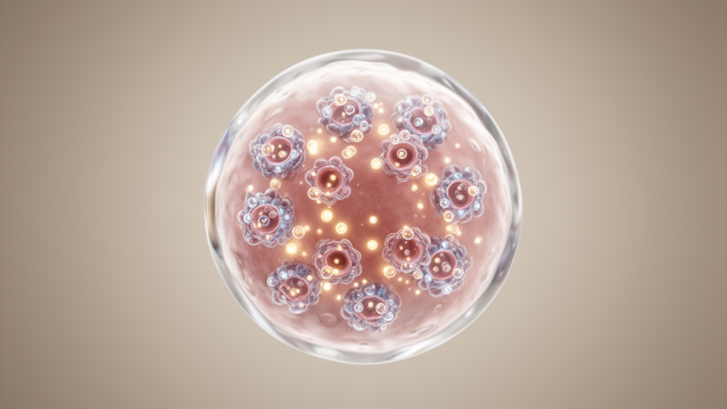
In a newly reported investigation, researchers have demonstrated for the first time that mature oocytes of the domestic cat functionally express the transient receptor potential vanilloid 3 channel, a calcium-permeable non-selective cation channel whose presence in feline gametes provides a molecular foundation for developing specialized assisted reproductive protocols. Intracellular calcium signaling serves as an indispensable trigger for oocyte maturation, egg activation, and fertilization across mammalian species, where repetitive calcium transients are required to convert a metaphase II arrested oocyte into a developing zygote. While prior work established that rodent oocytes rely on cation channels such as TRPV3 to mediate crucial calcium influx and divalent cation homeostasis, the expression and physiological operation of this channel in non-rodent carnivores remained uncharacterized. The research team combined transcript detection, immunocytochemical localization, live-cell calcium imaging, parthenogenetic cleavage assays, and comparative structural modeling to establish that feline oocytes express membrane-localized TRPV3 channels that respond to pharmacological agonists with robust cytosolic calcium elevations.
The study arrives against the backdrop of significant challenges in wildlife biology, where nearly half of all wild felid species worldwide currently face the threat of extinction. The International Union for Conservation of Nature lists fourteen felid species as vulnerable and four as endangered, including Chile’s native kodkod, which suffers from small and declining populations, severe habitat fragmentation, and direct persecution. Because collecting gametes directly from protected wild felids presents severe logistical and ethical hurdles, the domestic cat serves as the primary biomedical and reproductive model for developing assisted reproductive technologies. Techniques such as in vitro fertilization, intracytoplasmic sperm injection, and interspecific somatic cell nuclear transfer have generated live offspring in select wild felines by utilizing enucleated domestic cat oocytes as recipient cytoplasts. However, embryos produced via interspecific transfer frequently suffer from low developmental competence, substandard blastocyst formation, and compromised implantation rates, a deficiency largely attributed to an incomplete understanding of feline oocyte physiology and egg activation mechanisms.
Egg activation represents the pivotal signal transduction event during fertilization, initiated when sperm-oocyte fusion introduces sperm-specific phospholipase C zeta to induce periodic intracellular calcium oscillations. In experimental settings, artificial activation strategies attempt to mimic this physiological stimulus by promoting divalent cation influx, particularly calcium and strontium, through plasma membrane channels. In murine models, TRPV3 expression increases markedly during meiotic progression, peaking at the metaphase II stage, where its pharmacological stimulation triggers calcium entry sufficient to initiate parthenogenesis and block polyspermy through cortical granule exocytosis. Despite these established roles in rodents, existing activation protocols applied to domestic cat oocytes have predominantly been extrapolated from murine and bovine systems, yielding only modest success. The frequent occurrence of spontaneous activation during in vitro maturation and culture in feline gametes has further complicated protocol optimization, underscoring the urgent need to identify the endogenous channels governing calcium influx during feline meiosis.
To investigate feline gamete physiology, the researchers gathered sixty-two ovaries from thirty-one domestic cats undergoing routine ovariohysterectomies at regional veterinary clinics, isolating oocytes for controlled in vitro maturation. Morphological comparisons between mature feline and murine metaphase II eggs demonstrated striking interspecies structural divergences that bear direct physiological significance. Quantitative microscopy revealed that cat metaphase II eggs possess a mean cytoplasmic diameter of 137 plus or minus 1.7 micrometers, representing a statistically significant 1.8-fold increase compared to the 76.58 plus or minus 0.4 micrometers observed in mouse oocytes. Furthermore, feline eggs exhibited a dark, opaque cytoplasm resulting from substantial intracellular lipid storage, contrasting with the translucent cytoplasm of murine eggs. The zona pellucida surrounding feline eggs was also significantly thicker than that of mice, measuring 29.43 plus or minus 1.25 micrometers compared to 7.94 plus or minus 0.13 micrometers, representing an approximate 3.7-fold difference that emphasizes the distinct biomechanical environment of feline gametes.
Molecular analysis confirmed that the gene encoding TRPV3 is actively transcribed across key developmental stages of feline gametes. Using reverse transcription polymerase chain reaction with primers specifically designed against feline cDNA sequences retrieved from public genomic repositories, the researchers amplified a distinct 118-base-pair amplicon corresponding to feline Trpv3 from ovarian tissue, germinal vesicle oocytes, and in vitro matured metaphase II eggs. Amplification of the housekeeping control gene succinate dehydrogenase complex flavoprotein subunit A produced an expected 112-base-pair product across all examined samples. The consistent detection of target transcripts demonstrated that Trpv3 transcription is sustained throughout meiotic maturation, providing the cellular substrate necessary for channel synthesis before and after germinal vesicle breakdown.
Confocal immunofluorescence microscopy established the translation and spatial distribution of the TRPV3 protein in domestic cat oocytes across different meiotic stages. The primary antibody used in the assays was first validated in wild-type and TRPV3 knockout mouse eggs, where wild-type gametes exhibited clear peripheral membrane staining while knockout and secondary-antibody-only controls displayed no detectable fluorescence. In feline germinal vesicle oocytes, TRPV3 immunoreactivity was concentrated prominently along the cell periphery, co-localizing partially with cortical filamentous actin labeled by rhodamine phalloidin. As oocytes matured to the metaphase II stage, TRPV3 maintained strong enrichment at the plasma membrane, though cytoplasmic fluorescence intensity also increased noticeably. Fluorescence intensity profiling across oocyte cross-sections confirmed that signal intensity peaked sharply at the cell periphery and diminished toward the interior, illustrating that feline TRPV3 behaves as a membrane-associated channel that undergoes dynamic spatial redistribution during meiotic progression.
The functional competence of feline TRPV3 was verified through real-time ratiometric calcium imaging on zona pellucida-free metaphase II eggs loaded with the fluorophore Fura Red-AM. Confocal image acquisition monitoring excitation ratios at 405 and 488 nanometers revealed stable basal calcium levels under agonist-free control conditions. The application of the well-characterized TRPV3 agonist 2-aminoethoxydiphenyl borate elicited dose-dependent elevations in intracellular calcium concentration. While an agonist concentration of 100 micromolar produced a modest fluorescence ratio increase of 0.4319 plus or minus 0.1789 that did not reach statistical significance over baseline, the addition of 300 micromolar 2-APB induced a robust and statistically significant calcium response with a mean amplitude change of 0.6198 plus or minus 0.1585. These imaging recordings confirmed that pharmacological stimulation of feline TRPV3 reliably promotes extracellular calcium influx across the egg plasma membrane.
To evaluate whether TRPV3-mediated calcium entry could drive downstream developmental events, the team exposed mature metaphase II eggs to 300 micromolar 2-APB for one hour before culturing them in supplemented synthetic oviduct fluid for up to forty hours. Untreated control oocytes displayed an absence of spontaneous activation across all replicates, confirming the stability of the meiotic arrest under basal culture conditions. In contrast, eggs exposed to 300 micromolar 2-APB underwent parthenogenetic activation, achieving a mean cleavage rate of 39.97 plus or minus 22.93 percent at forty hours post-treatment. Although this demonstrated that TRPV3 stimulation generates sufficient calcium entry to overcome meiotic arrest and initiate early embryonic cleavage, the activation frequency was modest and required higher agonist concentrations and longer incubation periods than those reported for murine eggs, highlighting critical interspecies pharmacodynamic divergences.
To uncover the biophysical mechanisms underlying the reduced pharmacological sensitivity of feline TRPV3, the investigators carried out structural modeling and computational docking using Boltz-2, alongside binding pocket volume analysis using FPocket. Structural comparisons with the cryo-electron microscopy architecture of mouse TRPV3 revealed that while overall quaternary channel architecture is preserved, ligand-binding cavities differ substantially. Mouse TRPV3 features three distinct 2-APB binding pockets per subunit, situated within extracellular, transmembrane, and intracellular regions. In contrast, docking simulations on feline TRPV3 demonstrated that its intracellular pocket is constricted to a volume of 219.4 cubic angstroms, compared to 654 cubic angstroms in the mouse channel. Given the calculated molecular volume of 2-APB at approximately 192.1 cubic angstroms, the feline intracellular pocket is sterically incapable of accommodating the ligand, leaving the feline channel with only two functional binding sites per subunit.
These structural predictions aligned with calculated half-maximal effective concentrations determined across orthologs. In mouse TRPV3, predicted EC50 values for 2-APB were 0.26 micromolar at the extracellular site, 3.5 micromolar at the transmembrane site, and 11.02 micromolar at the intracellular cavity. For feline TRPV3, predicted EC50 values were 0.52 micromolar at the extracellular site and 3.4 micromolar at the transmembrane site, with no accessible intracellular site identified, while human ortholog values were 2.45 micromolar for transmembrane and 7.6 micromolar for intracellular sites. Comparative sequence alignments performed with MAFFT revealed that while mammalian TRPV3 orthologs share over ninety-two percent overall pairwise identity, feline TRPV3 harbors critical non-conservative substitutions in the pore-forming S6 transmembrane helix. Specifically, the mutations L596V, K614S, K616N, and A628S alter the lower activation gate, replacing the positively charged lysine cluster found in mice with neutral serine and asparagine residues, which likely eliminates stabilizing electrostatic interactions and modifies gate flexibility.
Beyond the pore gate, sequence alignments identified notable non-conservative substitutions in regulatory domains responsible for allosteric coupling and thermal sensitivity, including the ankyrin repeat domain, the S1 to S4 transmembrane bundle, the S4-S5 linker, and the TRP helix. Specific substitutions, such as Q114R, C131S, R136L, D198N, E207A, and K246S in the ankyrin repeats, along with S480G and the D468A mutation in the S4-S5 linker, directly impact channel mechanics. Because the S4-S5 linker functions as an essential mechanical gearbox that couples conformational movements in the sensory bundle to pore dilation, disrupting the electrostatic network via D468A provides a clear molecular rationale for the altered gating kinetics, modified thermal thresholds, and attenuated 2-APB sensitivity observed in feline eggs.
The identification of functional TRPV3 channels in feline oocytes provides valuable guidance for refining assisted reproductive technologies in endangered felids, while highlighting existing physiological limitations. Because 2-APB induces a monotonic calcium rise rather than the repetitive oscillations typical of natural mammalian fertilization, activating TRPV3 alone yields modest cleavage rates. Future artificial activation protocols for domestic cats and wild species such as the kodkod will likely require combining TRPV3 stimulation with agents that replicate physiologic oscillatory patterns, optimizing agonist concentrations specifically tailored to feline channel kinetics, or concurrently engaging complementary calcium-permeable pathways such as TRPM7 or Cav3.2 to maximize developmental competence.
Subject of Research: Functional expression and structural characterization of feline TRPV3 channels in oocytes
Article Title: Expression and functional characterization of the TRPV3 channel in domestic cat (Felis catus) mature oocytes
Article References: Vergara, S., Duarte-Zúñiga, J., Hidalgo, C., Suárez-Suárez, C., Castillo, K., Piesche, M., Aguilera, F., Fissore, R. A., Hinostroza, F., Veraguas-Dávila, D., & Carvacho, I. (2026). Expression and functional characterization of the TRPV3 channel in domestic cat (Felis catus) mature oocytes. Biological Research, 59(1), Article 53. https://doi.org/10.1186/s40659-026-00709-7
Image Credits: AI Generated
DOI: 10.1186/s40659-026-00709-7
Keywords: TRPV3, Felis catus, oocyte activation, calcium signaling, assisted reproductive technologies, ion channels, parthenogenesis, comparative biology, Expression, functional, characterization, channel
News Source: William Thompson. (November 7, 2026). Domestic Cat Oocytes Functionally Express Calcium-Permeable TRPV3 Ion Channels. Scienmag.