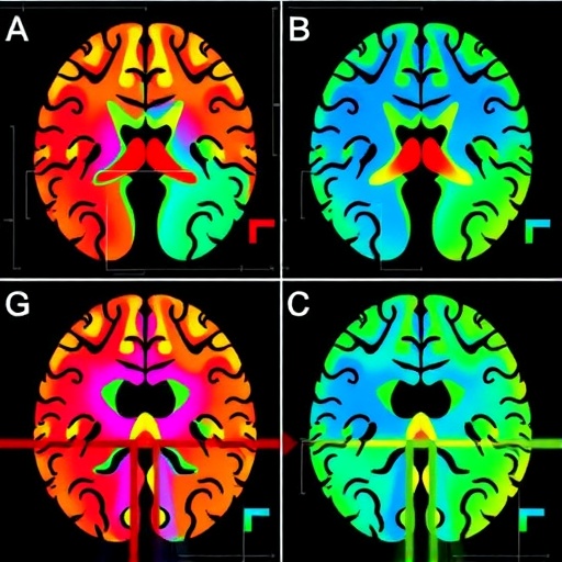
The hypothalamus, a thumbnail-sized structure deep in the brain that governs hunger, sleep, stress hormones and circadian rhythm, is emerging as an unexpected player in Alzheimer’s disease. A new study published in GeroScience reports that distinct hypothalamic regions shrink in different ways depending on whether a patient develops the disease early or late in life, and that the degree of shrinkage tracks not only with memory and cognition but also with mood disturbances that often accompany dementia. The findings, based on magnetic resonance imaging of living patients, add a rarely examined structure to the map of Alzheimer’s vulnerability and suggest that some of the disease’s most burdensome non-cognitive symptoms may have an anatomical anchor.
Alzheimer’s disease has long been classified into two broad clinical forms. Early-onset Alzheimer’s disease, which typically strikes before the age of 65, tends to produce more aggressive cognitive decline and affects posterior cortical regions such as the parietal and occipital lobes, where tau pathology accumulates excessively. Late-onset disease, the far more common form, follows a somewhat different anatomical script, with prominent involvement of medial temporal structures including the hippocampus. Yet patients with both forms frequently exhibit symptoms that cannot be explained by damage to memory circuits alone: depression, apathy, sleep disruption, weight loss and altered appetite. Neuroscientists have long suspected that the hypothalamus, the brain’s neuroendocrine control center, could be responsible for at least some of these deficits, but testing that idea in living patients has been technically difficult because the hypothalamus is small, irregularly shaped and sandwiched between fluid-filled spaces that confound standard brain-image analysis tools.
The research team, led by Giulia Quattrini of the IRCCS Istituto Centro San Giovanni di Dio Fatebenefratelli in Brescia, Italy, together with co-senior authors Marta Bortoletto and Moira Marizzoni and an international consortium of collaborators including Martina Bocchetta of University College London, tackled this problem using an automated segmentation method capable of parceling the hypothalamus into its component subunits on conventional T1-weighted MRI scans. The technique, developed in earlier work by the same imaging group, uses machine learning trained on expert-labeled data to divide the structure into anterior-superior, anterior-inferior, posterior, inferior and tubular regions, each with distinct cellular composition and neurochemical signaling profiles. This level of granularity matters because the hypothalamus is not a uniform blob: the anterior-superior sector houses neurons that promote wakefulness and regulate circadian timing, the posterior sector contains mammillary bodies critical for memory through their connections with the hippocampal fornix, and other subunits release vasopressin and oxytocin, hormones with documented roles in learning and emotional regulation.
The study population comprised 79 participants: 14 patients with early-onset Alzheimer’s disease, 28 with late-onset disease, 23 elderly healthy controls and 14 younger healthy controls. Diagnoses were made according to established clinical criteria from the National Institute on Aging and the Alzheimer’s Association, although the authors note that amyloid or fluid biomarker confirmation was not available for every participant, a limitation the team openly acknowledges. Each participant underwent structural MRI, and the resulting images were processed through the automated hypothalamic pipeline, with rigorous quality control of segmentation output. Beyond imaging, participants completed neuropsychological assessment of global cognition and memory, and clinical evaluation captured non-cognitive variables including depressive symptoms, measured with a geriatric depression scale, and body mass index, chosen because appetite dysregulation and weight loss are hallmark systemic features of advancing dementia and known correlates of hypothalamic function.
The volumetric results revealed a clear and asymmetric pattern. Patients with late-onset Alzheimer’s disease showed significantly reduced volumes of the whole hypothalamus and specifically its posterior region, bilaterally, compared with elderly healthy controls, with statistical thresholds corrected for multiple comparisons and significance levels reported at p less than 0.010. In contrast, early-onset patients did not differ significantly from controls in these posterior measures, suggesting that hypothalamic involvement is not a uniform feature of the disease but instead a signature that distinguishes the two clinical forms. One subunit, however, behaved differently: the right anterior-inferior region was smaller in patients relative to controls regardless of onset age, with a significance level of p equals 0.007, indicating that this sector may represent a common point of hypothalamic vulnerability across the Alzheimer’s spectrum. The anterior-inferior hypothalamus contains dense populations of neurons involved in autonomic and endocrine regulation, and its selective involvement in both patient groups hints at a shared pathophysiological process touching the neuroendocrine axis irrespective of disease onset.
Correlation analyses then connected structure to function. When patients with late-onset disease and elderly controls were pooled, smaller volumes of the altered hypothalamic regions were associated with worse global cognition, with Spearman correlation coefficients ranging from minus 0.35 to minus 0.30 and p values below 0.041. Memory performance also showed a positive association with hypothalamic integrity, with a correlation coefficient of 0.30 and p equals 0.041, a relationship that makes anatomical sense given the posterior hypothalamus’s mammillary bodies and their role in diencephalic memory circuits long recognized to be damaged in Alzheimer’s disease. Most striking, however, were the associations with mood: in the late-onset group, hypothalamic volumes correlated inversely with depressive symptom scores, with correlations as strong as rho equal to minus 0.82, significant at p below 0.031. In other words, patients with the most shrunken hypothalami reported the most severe depressive symptoms. Depression is one of the most common neuropsychiatric features of Alzheimer’s disease, affecting a substantial proportion of patients and accelerating functional decline, and previous research has linked late-life depression to dysregulation of the hypothalamic-pituitary-adrenal axis, the stress-hormone system whose central controller sits squarely within the hypothalamus.
The mechanistic plausibility of these findings is strengthened by decades of neuropathological work. Autopsy studies dating back to the classic Braak staging scheme have shown that tau pathology and neurofibrillary tangles can appear in the hypothalamus early in the disease process, sometimes preceding widespread cortical involvement. Tau and ubiquitin deposits have been documented in hypothalamic neurons of aged and Alzheimer’s disease brains, and amyloid-beta accumulation follows a predictable phase sequence that includes subcortical structures. Post-mortem investigations have further revealed profound degeneration of wake-promoting neurons in the hypothalamus of Alzheimer’s patients, providing a cellular substrate for the sleep and arousal disturbances that plague patients and caregivers alike. Animal studies add another dimension: pro-inflammatory interleukin-6 signaling in hypothalamic circuits has been shown to link cognitive impairment with peripheral metabolic alteration, and intranasal oxytocin as well as arginine vasopressin have each attenuated amyloid-induced memory deficits in rodent models, implicating hypothalamic neuropeptides as potential therapeutic agents rather than passive bystanders.
The metabolic connection is particularly compelling. Population-based autopsy data from the Hisayama study in Japan have demonstrated an association between hypothalamic Alzheimer’s pathology and body mass index, and independent imaging work has shown that hypothalamic volume correlates with body mass index in the general population. Weight loss in dementia is a serious clinical problem, associated with faster decline, increased frailty and higher mortality, and the current study’s inclusion of body mass index as a non-cognitive variable reflects a deliberate effort to capture this hypothalamic dimension of the disease, even if the imaging-to-metabolism correlations in this cohort did not dominate the statistical results. The authors emphasize that the hypothalamus should be viewed as both a culprit and a target in Alzheimer’s disease, a framing borrowed from earlier influential commentary, meaning that the structure may both contribute to systemic manifestations and suffer damage from the same pathological cascades that destroy cortical tissue.
The study’s design also carries implications for how researchers think about Alzheimer’s heterogeneity. The differential hypothalamic profile between early-onset and late-onset patients reinforces the growing view that these are not merely the same disease at different ages but partially distinct syndromes with different anatomical trajectories, genetic architectures and symptom clusters. Earlier work by the consortium and others has documented divergent cortical atrophy patterns, differing tau distribution and distinct cerebrospinal fluid biomarker profiles between the two forms. Adding the hypothalamus to this comparative framework opens the possibility that some of the clinical differences between early and late-onset disease, particularly in the non-cognitive realm, could be explained by differential subcortical involvement that has been largely invisible to conventional whole-brain volumetric analyses.
The authors are careful to frame their findings as hypothesis-generating rather than definitive. The sample sizes, while respectable for a study of a tiny brain structure, are modest, and the lack of universal biomarker confirmation means some diagnostic misclassification is possible. The cross-sectional design cannot establish whether hypothalamic atrophy precedes cognitive decline, accompanies it, or results from it, and longitudinal follow-up will be needed to determine whether hypothalamic volumes predict future symptom development or disease progression. Nonetheless, the study provides a technical demonstration that fine-grained hypothalamic volumetry is feasible in clinical research cohorts using widely available MRI sequences, and it flags specific subunits, the bilateral posterior hypothalamus and the right anterior-inferior region, as promising targets for larger, biomarker-anchored investigations. If future studies confirm and extend these results, hypothalamic imaging could eventually complement hippocampal measures in disease staging, and hypothalamically targeted interventions, from neuropeptide-based therapies to circadian and metabolic interventions, could enter the therapeutic conversation for a disease that still lacks treatments addressing its full clinical footprint, cognitive and non-cognitive alike. The study was approved by the local ethics committee and all participants provided written informed consent, and the dataset and analysis code are available from the corresponding authors on request.
Subject of Research: In vivo MRI-based volumetry of hypothalamic subregions in early- and late-onset Alzheimer’s disease and their associations with cognitive and non-cognitive symptoms
Subject of Research: Medicine
Article Title: Different hypothalamic regions in late-onset Alzheimer’s disease and aging: involvement and links with cognitive and non-cognitive features
Article References: Quattrini, G., Bocchetta, M., Bagattini, C., Saglia, S., Bertazzoli, G., Ferrari, E., Delai, M., Bulgari, M., Brignani, D., Canu, E., Agosta, F., Festari, C., Filippi, M., Gasparotti, R., Pievani, M., Cattaneo, A., Bortoletto, M., & Marizzoni, M. (2026). Different hypothalamic regions in late-onset Alzheimer’s disease and aging: involvement and links with cognitive and non-cognitive features. GeroScience. https://doi.org/10.1007/s11357-026-02488-1
Image Credits: AI Generated
DOI: 10.1007/s11357-026-02488-1
Keywords: Hypothalamus, Late-onset Alzheimer’s disease, Early-onset Alzheimer’s disease, MRI volumetry, Cognition, Memory, Mood, Depression, Body mass index, GeroScience
Cite Scienmag News
APA MLA Chicago
Cassandra Pierce. (September 9, 2026). Distinct hypothalamic regions implicated in late-onset Alzheimer’s disease and normal aging. Scienmag. https://scienmag.com/distinct-hypothalamic-regions-implicated-in-late-onset-alzheimers-disease-and-normal-aging/
Cassandra Pierce. “Distinct hypothalamic regions implicated in late-onset Alzheimer’s disease and normal aging.” Scienmag, 9 September 2026, https://scienmag.com/distinct-hypothalamic-regions-implicated-in-late-onset-alzheimers-disease-and-normal-aging/. Accessed 9 September 2026.
Cassandra Pierce. “Distinct hypothalamic regions implicated in late-onset Alzheimer’s disease and normal aging.” Scienmag. September 9, 2026. https://scienmag.com/distinct-hypothalamic-regions-implicated-in-late-onset-alzheimers-disease-and-normal-aging/
Copy citation Download RIS
Tags: Alzheimer’s disease brain vulnerabilityanatomical differences in Alzheimer’s diseasebrain regions associated with memory and cognitionbrain structure atrophy in agingbrain structures linked to Alzheimer’s pathologycircadian rhythm and stress hormone regulation in dementiacircadian rhythm disruption in dementiahypothalamic involvement in aginghypothalamic regions in Alzheimer’s diseasehypothalamic shrinkage and cognitive declinehypothalamus and brain aginghypothalamus and mood disturbanceshypothalamus and mood disturbances in dementiahypothalamus and neurodegenerationhypothalamus involvement in neurodegenerative diseaseslate-onset versus early-onset Alzheimer’sMRI brain imaging in dementiaMRI studies of hypothalamus in Alzheimer’sneuroanatomical markers of late-onneuroanatomy of Alzheimer’s diseasenon-cognitive symptoms of Alzheimer’s