


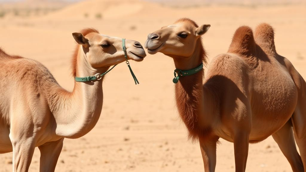
In the hyper-arid reaches of the Algerian Sahara, the dromedary camel has long been celebrated as a master of survival, capable of tolerating body water losses exceeding 25 percent of its total mass. A new cross-sectional study of 100 adult camels, published in BMC Zoology, now reports that severe dehydration is associated with a striking physical contraction of the spleen’s marginal zone, a lymphoid compartment that serves as the first line of defense against blood-borne pathogens. In dehydrated animals, the marginal zone was on average 13.7 percent thinner than in hydrated controls, and the density of B cells packed within that zone fell by roughly 24 percent.
The research team, working in the El Oued region and the transitional steppe zone around Bir El Ater roughly 200 kilometers to the north, sampled camels at municipal abattoirs between 2023 and 2025. All animals were clinically healthy adults aged 4 to 15 years, non-pregnant and non-lactating females, selected only after passing rigorous ante-mortem and post-mortem inspections by official state veterinarians. Animals showing generalized lymphadenopathy, visceral parasitic cysts, signs of trypanosomiasis or tuberculosis, or any lesions warranting carcass condemnation were strictly excluded, a filter designed to minimize the confounding effects of subclinical infection and parasite burden.
Hydration status, the study’s primary exposure variable, was defined physiologically rather than assumed. Blood samples taken by jugular venipuncture were analyzed for serum osmolality by freezing-point depression osmometry and for hematocrit by micro-capillary centrifugation. Because euhydrated camels typically maintain serum osmolality between 290 and 300 mOsm/kg, animals exceeding 310 mOsm/kg were classified as dehydrated, with elevated packed cell volume values corroborating the classification. This yielded 58 hydrated and 42 dehydrated animals. The physiological validation was unambiguous: dehydrated camels showed a mean serum osmolality increase of 49.7 mOsm/kg and an 11.6 percent rise in hematocrit, and the two markers correlated strongly with each other, with a coefficient of 0.81.
To quantify the splenic changes, the researchers harvested spleen samples within 45 minutes of slaughter and excised tissue blocks from a standardized location on the mid-parietal surface, cut perpendicularly to the capsule so every block captured the full architectural continuum from capsule to deep white pulp. After fixation and paraffin embedding, serial sections were stained with hematoxylin and eosin, and B cells were labeled by immunohistochemistry using a rabbit anti-CD20 polyclonal antibody previously validated for camel species. Digital morphometry was performed in QuPath and ImageJ by investigators blinded to both hydration status and regional origin, and a subset of 20 slides independently re-evaluated by a second blinded observer showed excellent agreement, with an intraclass correlation coefficient of 0.91.
The morphometric results were pronounced. Mean marginal zone thickness fell from 183.1 plus or minus 9.6 micrometers in hydrated controls to 157.9 plus or minus 10.8 micrometers in dehydrated animals, a difference the authors report as highly significant. A sensitivity analysis treating serum osmolality as a continuous predictor confirmed a significant negative linear association with marginal zone thickness. Distributional analysis revealed what the authors describe as a bimodal compression, a shift toward a narrower structural baseline suggesting that dehydration may impose a physiological constraint overriding natural morphological variation. Marginal zone thickness also correlated negatively with serum osmolality and hematocrit, and positively with body condition score, hinting that energetic reserves may buffer against the atrophy.
Parallel immunohistochemical quantification showed that B-cell density within the marginal zone dropped from 528.8 plus or minus 103.4 cells per square millimeter in hydrated animals to 400.4 plus or minus 95.1 cells per square millimeter in the dehydrated cohort. Regression modeling indicated that marginal zone thickness was a robust positive predictor of B-cell density, explaining 59 percent of the variance, a pattern the authors interpret as evidence that the physical collapse of the compartment constrains the niche space available for B-lymphocyte retention. Importantly, the authors frame this as a statistical association rather than proof of a causal sequence.
To probe that structure-cell relationship further, the team applied a formal mediation analysis with bootstrap resampling. Within the specified statistical model, the pathway running from dehydration status through marginal zone thickness to B-cell density, estimated at an average causal mediation effect of minus 196.2 cells per square millimeter, accounted for approximately 59 percent of the total dehydration-B-cell association of minus 332.5 cells per square millimeter. The authors are explicit that this proportion mediated is a statistical decomposition, not evidence that dehydration physically collapses the marginal zone or that such collapse causes B-cell loss. Because all three variables were measured at a single time point in a cross-sectional design, temporal ordering cannot be established, and the assumptions required for causal mediation cannot be fully verified.
The study also uncovered a geographic gradient in splenic architecture. Camels from the hyper-arid Saharan site at El Oued showed greater marginal zone contraction than those from the semi-arid steppe at Bir El Ater, with a 15.9 percent reduction in thickness in the Saharan cohort compared with a milder 10.6 percent reduction in the steppe population. Notably, Saharan camels possessed a significantly thinner marginal zone even when hydrated, at 176.4 versus 188.5 micrometers. The authors hypothesize this thinner baseline phenotype may reflect chronic eco-physiological acclimatization to a resource-scarce environment, but they caution that lifelong differences in forage quality, nutritional history, and management conditions between the regions cannot be excluded as primary drivers, and that regional contrasts alone cannot establish adaptation.
The authors are equally careful about what the findings do not show. No functional immune assay was performed, so the reduced B-cell density could be transient, detrimental, neutral, or even beneficial during dehydration; the data cannot distinguish among these possibilities. The remodeling may reflect hemoconcentration, altered perfusion, endocrine stress, altered energy balance, or nutritional status rather than a dehydration-specific response. Cortisol and other endocrine stress markers were not measured, precise information on time since last watering, transport duration, and nutritional history was unavailable, and minor subclinical infections could not be absolutely excluded. The proposed hemodynamic mechanism, in which rising blood viscosity alters perfusion through the intricate marginal sinusoids of an organ uniquely equipped with an extensive subcapsular sinus system, remains explicitly untested because no microvascular endothelial staining or blood flow measurements were undertaken.
What the study does deliver is the first quantitative, organ-level link between systemic hydration status and splenic microarchitecture in free-ranging dromedaries, filling a gap in a literature that had described gross anatomy and hematological parameters in isolation. The splenic marginal zone houses the specialized B cells responsible for rapid neutralization of blood-borne pathogens, making it a compartment of real immunological interest in an animal whose spleen also doubles as a capacious erythrocyte reservoir. The authors suggest the reduction in B-cell density could be associated with altered splenic immune capacity, while noting this remains unverified. Future longitudinal or controlled dehydration-rehydration studies incorporating endocrine, metabolic, nutritional, inflammatory, and microvascular measurements, they conclude, will be needed to determine whether the remodeling is reversible, specific to hydric stress, and functionally consequential for host defense.
Subject of Research: Dehydration-associated remodeling of the splenic marginal zone and B-cell depletion in the dromedary camel
Article Title: Splenic plasticity at the arid limit: dehydration is associated with microanatomical remodelling and B-cell depletion in the dromedary camel (Camelus dromedarius)
Article References: Fares, M. A., Mouane, A., Elmounedi, N., Mlik, R., Chikha, M., Ferag, A., Messaoudi, M., Atoki, A. V., & Faye, B. (2026). Splenic plasticity at the arid limit: dehydration is associated with microanatomical remodelling and B-cell depletion in the dromedary camel (Camelus dromedarius). BMC Zoology, 11(1), Article 28. https://doi.org/10.1186/s40850-026-00284-0
Image Credits: AI Generated
DOI: 10.1186/s40850-026-00284-0
Keywords: dromedary camel, spleen, marginal zone, dehydration, B cells, immunohistochemistry, serum osmolality, Sahara, histomorphometry, mediation analysis, BMC Zoology, desert physiology
News Source: Drew Townsend. (October 29, 2026). Dehydration thins the spleen’s immune hub in desert camels. Scienmag.



