Researchers at the Medical University of South Carolina identify mutations in ciliary genes causing bicuspid aortic valve — the most common heart valve birth defect — and aortic valve narrowing

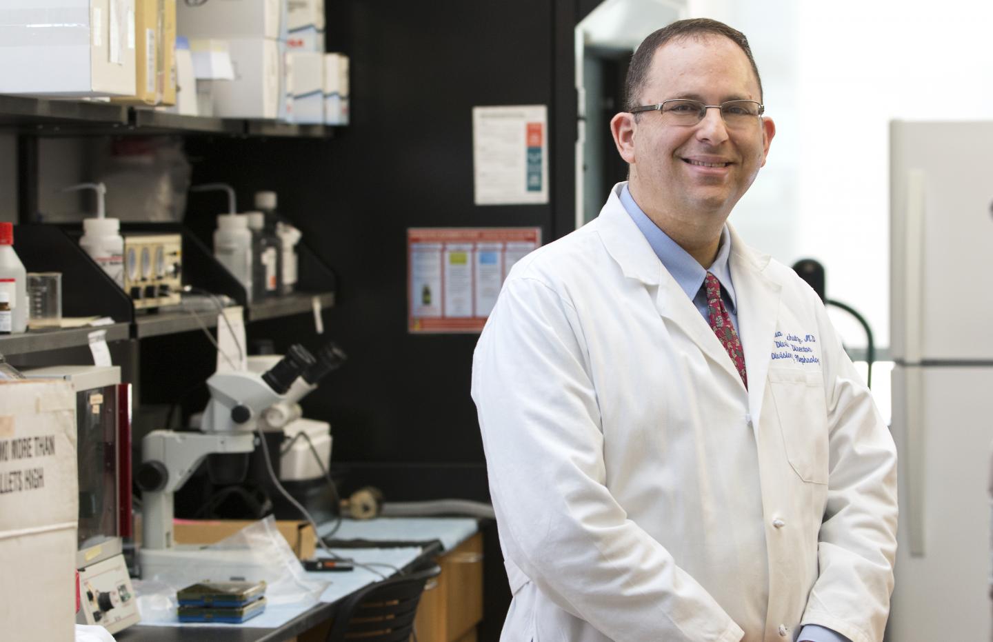
Credit: Sarah Pack, Medical University of South Carolina
Bicuspid aortic valve (BAV) is the most common heart valve birth defect and one of the most common birth defects of any type, affecting around 70 million people worldwide. A healthy aortic valve has three leaflets; in BAV disease, two of the leaflets are fused together, impairing the function of the valve. In many individuals with BAV, the valves eventually will have to be replaced or repaired through heart surgery.
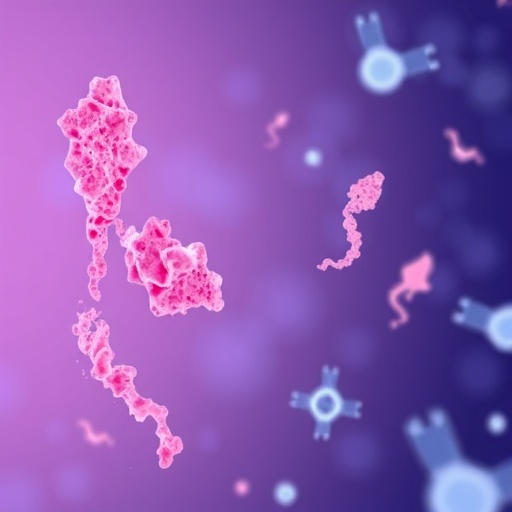
A team of researchers at the Medical University of South Carolina (MUSC) have discovered that a mutation in a gene controlling the production of cilia, tiny antennae protruding from the cell surface, are linked to the development of BAV.
In an article published online August 7, 2019 in the journal Circulation, the team used animal models and human data to reveal that BAV disease and aortic valve narrowing are caused by disruption of the exocyst, a shuttling complex that moves cilia cargo to the cell membrane and allows for the development of cilia, and defects in cilia production.
Most cells have cilia, which help them “sense” their surroundings.
“You can think of cilia like tiny antennae that cells use to transmit information to each other,” said Diana Fulmer, Ph.D. candidate at MUSC and lead author of the manuscript. “In heart valves, cilia function as signaling hubs during development to coordinate how cells and extracellular matrix arrange to form mature tissue.”
Only recently has the medical community begun to realize how cilia are involved in human health and disease.
“Up until 20 years ago, people were writing that cilia were vestigial organelles and had no function,” said Joshua H. Lipschutz, M.D., professor of medicine and director of the Division of Nephrology at MUSC and co-senior author of the article. “Now, there’s a whole field of ciliopathies.”
Russell A. Norris, Ph.D., associate professor of medicine in the Department of Regenerative Medicine and Cell Biology at MUSC and an expert in heart valve biology and genetics, is co-senior author of the manuscript. Norris first started to suspect that cilia were involved in BAV disease when he noticed that patients with certain ciliopathies, or cilia diseases, also had BAV disease. This observation led to a collaboration with Lipschutz, who studies autosomal dominant polycystic kidney disease, or ADPKD, which is a cilia disease affecting the kidney.
“We knew that ADPKD patients have a much higher incidence of heart defects, including bicuspid aortic valve disease,” said Lipschutz. “So, there was a very strong clinical association.”
Norris and Lipschutz wrote a collaborative grant with Simon Body, M.D., director of the Bicuspid Aortic Valve Consortium and an associate professor of anaesthesia at Harvard Medical School, to find out how cilia contribute to BAV disease. “It was really a true collaboration,” said Lipschutz. The grant was funded through the American Heart Association.
The investigators first compared the genomes of healthy adults and adults with BAV disease. In a genome-wide association study, the most associated differences in genomes were found in or near genes that are important in regulating ciliogenesis through the exocyst.
Animal models were then used to determine whether mutations in a central exocyst protein, Exoc5, caused disease in other organisms. Knocking out or “disabling” the gene for Exoc5 in zebrafish severely obstructed blood flow from the heart, leading to poor cardiac function and early death. The zebrafish also showed signs of other ciliopathies.
Remarkably, injecting the correct gene sequence for Exoc5 into the zebrafish prevented them from developing heart defects, proving that the mutation was causing the disease.
The investigators then disabled the Exoc5 gene in mice, specifically in the cells that make up the cardiac valves. Mice with both copies of the disabled gene died early during development. Mice with only one copy of the disabled gene were born but had a high rate of BAV disease and problems with cilia production.
Humans with BAV disease often develop calcification on their aortic valve, leading to stenosis, or narrowing, of the valve opening. This can cause severe functional defects within the heart and is a major indication for surgery.
Interestingly, mice with the disabled exocyst gene also developed calcified aortic valves and had significantly larger aortic roots.
“We saw the same thing we see in people,” said Lipschutz. “This gives us a good animal model for the disease.”
This study provides novel insight into the origin of BAV disease.
“We showed that the cause of a very common, potentially lethal, genetic defect in people was due to cilia and the exocyst,” Lipschutz said.
Knowledge gained from the study should accelerate the development of new therapies.
“Finding the cause for disease is the first step to finding a cure,” said Lipschutz. “Now that we know what’s causing it, we can come up with ways to treat it without surgery.”
###
About MUSC
Founded in 1824 in Charleston, MUSC is the oldest medical school in the South, as well as the state’s only integrated, academic health sciences center with a unique charge to serve the state through education, research and patient care. Each year, MUSC educates and trains more than 3,000 students and 700 residents in six colleges: Dental Medicine, Graduate Studies, Health Professions, Medicine, Nursing and Pharmacy. The state’s leader in obtaining biomedical research funds, in fiscal year 2018, MUSC set a new high, bringing in more than $276.5 million. For information on academic programs, visit http://musc.
As the clinical health system of the Medical University of South Carolina, MUSC Health is dedicated to delivering the highest quality patient care available, while training generations of competent, compassionate health care providers to serve the people of South Carolina and beyond. Comprising some 1,600 beds, more than 100 outreach sites, the MUSC College of Medicine, the physicians’ practice plan, and nearly 275 telehealth locations, MUSC Health owns and operates eight hospitals situated in Charleston, Chester, Florence, Lancaster and Marion counties. In 2019, for the fifth consecutive year, U.S. News & World Report named MUSC Health the number one hospital in South Carolina. To learn more about clinical patient services, visit http://muschealth.
MUSC and its affiliates have collective annual budgets of $3 billion. The more than 17,000 MUSC team members include world-class faculty, physicians, specialty providers and scientists who deliver groundbreaking education, research, technology and patient care.
Media Contact
Heather Woolwine
[email protected]
Related Journal Article
http://dx.


