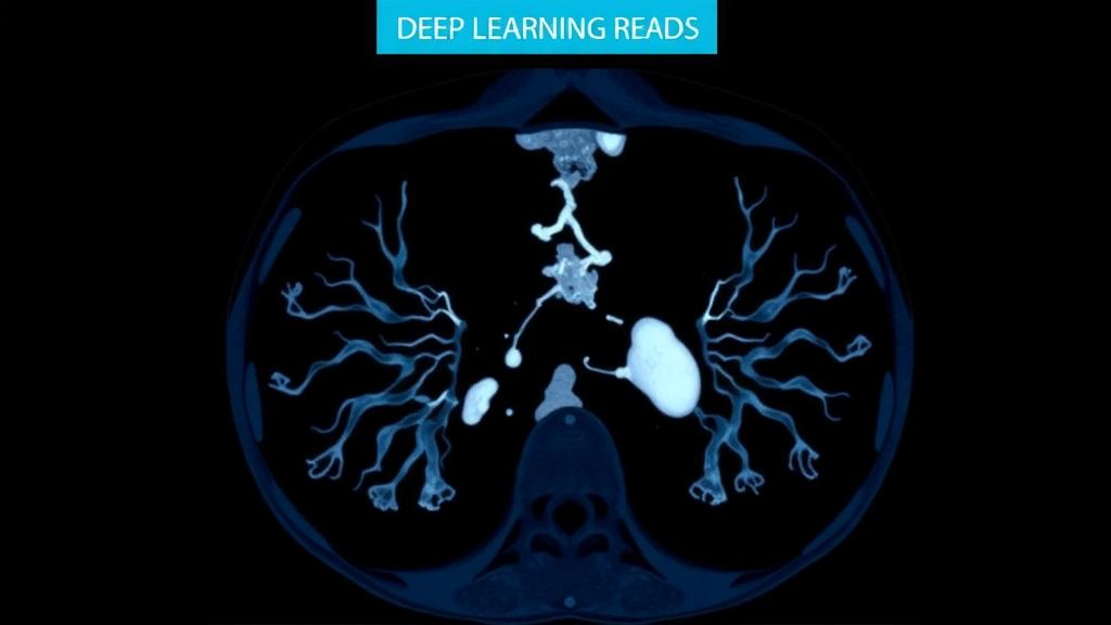
When a patient arrives in an intensive care unit with coronavirus disease, one of the most urgent questions facing clinicians is deceptively simple: is the infection getting worse or getting better? The answer shapes oxygen strategies, antiviral decisions, isolation measures and the timing of escalation to more aggressive support. Yet in practice, determining whether a patient sits in the progression or remission stage of COVID-19 has relied on a patchwork of laboratory values, clinical scores and radiologists’ visual readings of chest scans, none of which offers a rapid and reliable verdict on its own. A new study published in BMC Medical Imaging by a team of clinicians and computer scientists in Nanjing, China, argues that a purpose-built artificial intelligence system can close that gap, reading three-dimensional chest computed tomography scans to stage the disease with accuracy that outperforms manual interpretation in their setting.
The system, named COV-DSNet, was developed jointly by researchers from the Department of Critical Care Medicine at Nanjing Drum Tower Hospital, The Affiliated Hospital of Nanjing University Medical School, and the National Key Laboratory for Novel Software Technology at Nanjing University. It belongs to a family of algorithms known as three-dimensional convolutional neural networks, which process volumetric medical images as complete spatial objects rather than as stacks of independent two-dimensional slices. This distinction matters for pneumonia imaging, because the lesions of COVID-19 — ground-glass opacities, consolidations and the characteristic peripheral distributions of inflamed lung tissue — extend across many slices, and their overall burden and evolution are inherently volumetric phenomena.
Architecturally, COV-DSNet combines two ideas that have become central to modern medical image analysis. The first is mixed convolution, in which the network applies three-dimensional convolutional kernels to some layers and two-dimensional kernels to others, balancing the need to capture spatial context across the lung volume against the need to extract fine-grained textural features within individual slices. The second is the incorporation of attention mechanisms, computational modules that allow the network to learn which regions of an image deserve greater weight when making a decision. In effect, attention steers the model toward the diseased portions of the lung and away from irrelevant structures such as the heart, mediastinum or imaging artifacts, mimicking in a limited way the focused reading strategy of an experienced radiologist.
To train and evaluate the system, the team drew on data from 578 patients with COVID-19, combining pulmonary CT scans with clinical data screening. After applying their inclusion criteria, 297 CT scans from 156 patients entered the COV-DSNet analysis. The task posed to the network was binary staging: given a scan, classify the patient as being in the progression stage or the remission stage of the disease. This framing reflects the clinical reality that the two stages call for different management priorities — progression demands anticipation of deterioration and closer monitoring, while remission supports de-escalation and planning for discharge.
The performance figures reported in the study are grounded in a standard evaluation metric known as the area under the receiver operating characteristic curve, or AUC-ROC, which summarizes a model’s ability to discriminate between the two classes across all possible decision thresholds. Across their analyses, COV-DSNet achieved a mean AUC-ROC of 0.820, with a standard deviation of 0.030 and a 95 percent confidence interval spanning 0.792 to 0.844. In a randomly assigned verification set, the system reached an AUC-ROC of 0.864, with a 95 percent confidence interval of 0.750 to 0.947, alongside a sensitivity of 0.708 and a specificity of 0.921. The authors note that this specificity — the proportion of patients correctly identified as not being in progression — was higher than that of manual reading under the pre-specified single-center scope and reader setting of the study.
That emphasis on specificity is clinically meaningful. A staging tool that frequently raises false alarms would trigger unnecessary interventions, prolonged isolation and wasted resources, eroding the trust of the clinicians who are meant to use it. A sensitivity of 0.708 means the system missed roughly three in ten progression cases in the verification set, a figure the authors present transparently, but the high specificity suggests that when COV-DSNet declares a patient to be in progression, that judgment is usually warranted. In a triage context, such a signal could help prioritize which patients receive the closest attention during a surge of admissions.
Perhaps the most consequential finding of the study concerns what the AI-derived stage adds to conventional prognostic modeling. The researchers built a baseline clinical model using two established measures: the APACHE II score, a widely used severity-of-illness metric computed from physiological measurements and age, and the PaO2/FiO2 ratio, which quantifies how efficiently oxygen passes from the lungs into the blood. On its own, this clinical model predicted severe progression within seven days with an AUC of 0.908. When the COV-DSNet-derived stage diagnosis was added, the AUC rose to 0.978 — a substantial improvement in a prediction task that directly informs decisions about intensive monitoring and early escalation of care. The combined model also showed good calibration, with a bootstrap-corrected mean absolute error of 0.047, meaning its predicted probabilities tracked closely with observed outcomes rather than being systematically overconfident or underconfident.
The study also explored a second, less obvious application: infectivity assessment. Reverse transcription-polymerase chain reaction tests, the standard for detecting SARS-CoV-2, report a cycle threshold, or Ct value, which reflects how much viral genetic material is present in a sample. Lower Ct values generally indicate higher viral loads, and Ct values have been used to guide judgments about how infectious a patient is and how long isolation should continue. The researchers found that in a subset of COVID-19 patients, the AI-derived staging helped avoid misjudgments about infectivity that would have arisen from relying on the RT-PCR Ct value alone. This suggests that the structural state of the lungs, as captured by CT, carries information about the disease process that viral load measurements do not fully encompass, and that integrating imaging-based staging could refine infection control decisions at the bedside.
The authors are careful to frame their conclusions within the limits of the data. The study was conducted at a single center, Nanjing Drum Tower Hospital, and the comparison with manual reading was made under a pre-specified reader setting, so the reported advantage over human interpretation may not generalize to every clinical environment or every level of radiological expertise. The verification set was modest in size, as reflected in the width of its confidence interval, and the work was carried out on patients from a single institution’s population. External validation on independent cohorts from other hospitals and regions remains a necessary step before any claim of broad clinical readiness, and the authors position the system as a rapid staging tool demonstrated within their single-center study rather than as a finished diagnostic product.
Even with those caveats, the work points toward a broader role for volumetric deep learning in the management of respiratory infections. The authors suggest that their approach provides new ideas and solutions for the early clinical management of other pneumonias beyond COVID-19, since the core problem — inferring disease phase from the evolving appearance of lung tissue on CT — is not unique to SARS-CoV-2. If staging algorithms of this kind can be validated across institutions, they could become a routine layer of decision support, converting the raw three-dimensional information in a chest scan into an actionable estimate of where a patient stands in the arc of their illness. For clinicians confronting the next wave of severe respiratory disease, that could mean the difference between reacting to deterioration after it happens and anticipating it days in advance.
Subject of Research: Deep learning-based staging of COVID-19 using chest CT scans
Article Title: Enhanced coronavirus disease staging with deep learning of chest computed tomography scan images
Article References: Chen, M., Cai, H., Lu, H., Ling, T., Gao, Y., You, Y., Wang, Y., Cao, K., Shi, Y., Zhang, J., & Yu, W. (2026). Enhanced coronavirus disease staging with deep learning of chest computed tomography scan images. BMC Medical Imaging. https://doi.org/10.1186/s12880-026-02825-y
Image Credits: AI Generated
DOI: 10.1186/s12880-026-02825-y
Keywords: COVID-19, deep learning, chest CT, 3D convolutional neural network, attention mechanisms, disease staging, artificial intelligence, BMC Medical Imaging, AUC-ROC, APACHE II, RT-PCR Ct value, pneumonia
News Source: Blake Davidson. (October 8, 2026). Deep Learning Reads Chest CT Scans to Stage COVID-19 Patients Faster. Scienmag.