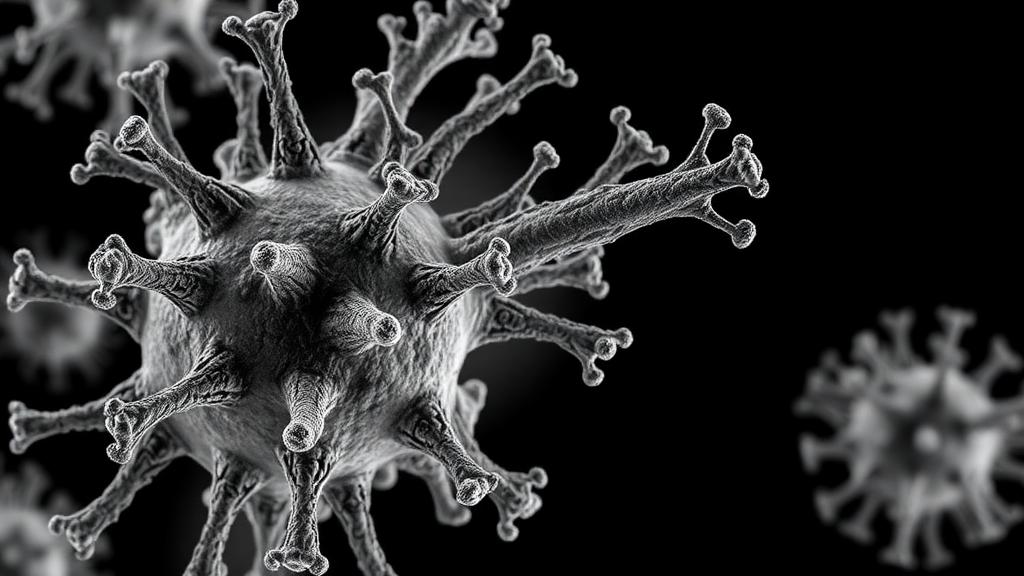
Bacteria are locked in a ceaseless evolutionary war with bacteriophages, the viruses that prey on them, and one of their most intriguing weapons in this conflict has just come into sharp molecular focus. A team of researchers at the Indian Institute of Science in Bengaluru has determined the first three-dimensional structure of a TenpIN toxin-antitoxin system, a bacterial anti-phage defense module that is shared, in identical form, by several major pathogenic species including Salmonella enterica, Shigella flexneri, Klebsiella pneumoniae, and Escherichia coli. Using single-particle cryo-electron microscopy, the scientists resolved the complex at approximately 3.9 angstrom resolution, revealing an elegant ring-like machine built from just four molecules: two copies of a protein toxin called TenpN and two copies of a non-coding RNA antitoxin called tenpI.
Toxin-antitoxin systems are genetic modules widespread among bacteria and archaea that pair a growth-inhibiting toxin with a cognate antitoxin that keeps it in check. In type III systems, the antitoxin is not a protein but an RNA molecule that directly binds and neutralizes the toxin. Under normal conditions the pair sits in a dormant embrace, but when a phage attacks, the antitoxin is overwhelmed or neutralized, freeing the toxin. The unleashed toxin acts as an endoribonuclease, cleaving both bacterial and phage RNA and thereby halting viral replication in an act of altruistic suicide that protects the wider bacterial population from infection.
Type III toxin-antitoxin systems have traditionally been divided into three families based on toxin sequence homology: toxIN, cptIN, and tenpIN. While the ribonucleoprotein complexes of the ToxIN and CptIN systems had already been structurally characterized, the tenpIN family remained the least understood member of the trio, with no structure available since its first functional characterization in the bacterium Photorhabdus luminescens more than a decade ago. Recent bioinformatic surveys have changed the picture dramatically, identifying more than 700 unique tenpN sequences across prokaryotic genomes and viruses, and revealing that these systems are far more widespread than previously appreciated, including within the ESKAPE pathogens responsible for most antibiotic-resistant infections.
The research team selected a putative tenpIN system whose toxin sequence is 100 percent identical across several enterobacterial pathogens, making it an attractive representative for structural study. The genetic architecture follows the canonical type III layout: a shared promoter drives an antitoxin cassette of 102 nucleotides, followed by a Rho-independent terminator and a toxin gene encoding a 153-amino-acid protein. To confirm the system’s function, the researchers expressed the TenpN toxin alone in E. coli cells and observed significant growth inhibition, a bacteriostatic effect that was fully rescued when the tenpI antitoxin sequence was co-expressed. These assays established that the conserved pathogenic sequence encodes a genuine, working type III toxin-antitoxin module.
A striking feature of this system emerged from close inspection of the antitoxin sequence. Standard tandem-repeat-finding software could not detect any repeats at all, because unlike the near-identical repeats of toxIN and cptIN systems, the tenpI antitoxin contains two non-identical repeats, termed Rp1 and Rp2, which differ at multiple nucleotide positions, and Rp2 is three nucleotides shorter than Rp1. Despite this sequence divergence, both repeats were predicted to fold into H-type pseudoknots, the hallmark tertiary structure of type III antitoxin RNAs. When each repeat was cloned separately and tested, both Rp1 and Rp2 independently rescued bacteria from TenpN-mediated growth inhibition, and colony-forming unit assays showed that the full-length antitoxin, Rp1, and Rp2 all counteracted toxicity to a similar extent.
To visualize the complex, the team co-expressed and co-purified the toxin and antitoxin from E. coli using affinity, ion-exchange, and size-exclusion chromatography. The purified complex eluted at an apparent molecular weight of roughly 72 kilodaltons, consistent with a heterotetramer of two toxin proteins and two antitoxin RNA molecules, while free TenpN eluted as an 18-kilodalton monomer. Sequencing of the RNA recovered from the purified complex confirmed that both Rp1 and Rp2 were incorporated. Circular dichroism thermal melting experiments showed that both repeats unfold with a melting temperature of about 56 degrees Celsius, demonstrating that the non-identical repeats are equally thermally stable, and in vitro assembly experiments confirmed that TenpN forms complexes with either repeat on its own.
The cryo-EM analysis itself demanded careful sample preparation. Negative-stain transmission electron microscopy identified the most homogeneous particle fractions, and cryo-EM data collected on a 200-kilovolt Talos Arctica microscope with a K2 direct electron detector yielded 2,855 movies. From more than a million automatically picked particles, iterative two-dimensional and three-dimensional classification converged on a well-resolved reconstruction built from 327,240 particles. The final map displayed clear C2 symmetry, and symmetric and asymmetric reconstructions proved nearly identical, indicating no significant structural asymmetry within the assembly. The resulting structure, determined in the complex’s native state without antibodies or tags, measures approximately 80 by 60 angstroms and shows the protein and RNA components arranged alternately in a closed cyclic ring with a hole at its center.
Model building combined computational prediction with experimental density. An AlphaFold2 model of TenpN was fitted and refined into the map, while the RNA had to be constructed de novo, fragment by fragment, using tools including simRNA, RhoFold+, Coot, Chimera, and Phenix. The Rp1 model fit the density better than Rp2, reflected in map-to-model correlation coefficients of 0.66 versus 0.52, likely because Rp2 lacks three nucleotides, so Rp1 was used to represent both RNA positions. The refined structure shows TenpN as a well-folded protein of six alpha-helices and five beta-strands, with a four-stranded antiparallel beta-sheet core and a long kinked helix that closely mirrors features previously seen in ToxN, where the kinked helix helps hold the antitoxin RNA in place. The tenpI RNA folds into a central H-type pseudoknot with two stems and two loops, its single-stranded 5-prime and 3-prime ends binding along an electropositive groove on the toxin surface in a head-to-tail arrangement that generates the cyclic tetramer.
Comparative analysis placed TenpN firmly within the type III toxin lineage. Despite sharing only about 13 percent sequence identity with ToxN and CptN, TenpN adopts a conserved overall fold, and DALI structural comparison showed it resembles ToxN, with a backbone root-mean-square deviation of 5.4 angstroms, more closely than CptN, at 10.1 angstroms. Sequence alignment highlighted conserved residues in the region forming the catalytic center, prompting the researchers to create alanine mutants of TenpN residues Pro53, Leu54, Arg55, and Ser56. Growth assays revealed that P53A and L54A mutants remained fully toxic, while R55A and S56A lost their ability to inhibit bacterial growth, identifying Arg55 and Ser56 as functionally critical residues that likely constitute the putative active site of the TenpN endoribonuclease, analogous to the validated active-site triads of ToxN and CptN.
The structural comparison also revealed a fascinating chimeric character of the TenpIN system: the toxin resembles ToxN, yet the antitoxin RNA is architecturally closer to cptI. Both tenpI and cptI feature a single-nucleotide loop that twists to coaxially stack the second stem beneath the first, and a long A-rich second loop that runs behind the first stem like a third strand, potentially forming A-minor interactions, whereas toxI RNA has shorter stems, more balanced loops, and a distinctive UUU triplet at the helical junction. These RNA size differences appear to dictate assembly stoichiometry: the larger cptI and tenpI RNAs form heterotetramers with their toxins, while the compact toxI assembles a heterohexamer. Because the single-stranded ends of tenpI and cptI converge on the same side of the active site, the tetrameric arrangement follows naturally. Beyond resolving an evolutionary puzzle, the structure opens a practical avenue: small molecules or peptides designed to disrupt the TenpIN complex could free the toxin inside pathogenic bacteria, offering a fundamentally new antibacterial strategy at a time when resistance to conventional antibiotics continues to spread.
Subject of Research: Cryo-EM structure of the TenpIN type III toxin-antitoxin ribonucleoprotein complex in pathogenic bacteria
Article Title: Cryo-EM reveals the quaternary architecture of TenpIN type III toxin-antitoxin RNP complex common to pathogenic bacteria
Article References: Nadig, K., Padmanaban, S., Noor, S., Sandhya, S., Manikandan, P., Dutta, S., & Singh, M. (2026). Cryo-EM reveals the quaternary architecture of TenpIN type III toxin-antitoxin RNP complex common to pathogenic bacteria. PLOS Pathogens, 22(10), e1014690. https://doi.org/10.1371/journal.ppat.1014690
Image Credits: AI Generated
DOI: 10.1371/journal.ppat.1014690
Keywords: cryo-EM, toxin-antitoxin systems, TenpIN, bacteriophage defense, RNA pseudoknot, endoribonuclease, pathogenic bacteria, ESKAPE pathogens, structural biology, antimicrobial targets, ribonucleoprotein complex, bacterial immunity
News Source: Kristina Jarvis. (October 10, 2026). Cryo-EM Exposes Hidden Architecture of a Bacterial Antiviral Weapon Found in Pathogens. Scienmag.