A thin, transparent window at the front of the eye may be quietly reporting on the state of the nervous system far beyond it. A narrative review published in the British Journal of Biomedical Science argues that the cornea’s dense meshwork of sensory nerves, imaged non-invasively with in vivo confocal microscopy (IVCM), could serve as an adjunctive biomarker for a wide range of neurodegenerative and neurological conditions, from diabetic peripheral neuropathy to Parkinson’s disease, dementia, multiple sclerosis and beyond. The authors, led by Jiayue Li and Andrew En Hsien Sim of the Yong Loo Lin School of Medicine at the National University of Singapore, stress that the evidence is far stronger for some diseases than for others, and that corneal nerve measurements are best seen today as promising adjuncts rather than established diagnostic tools.
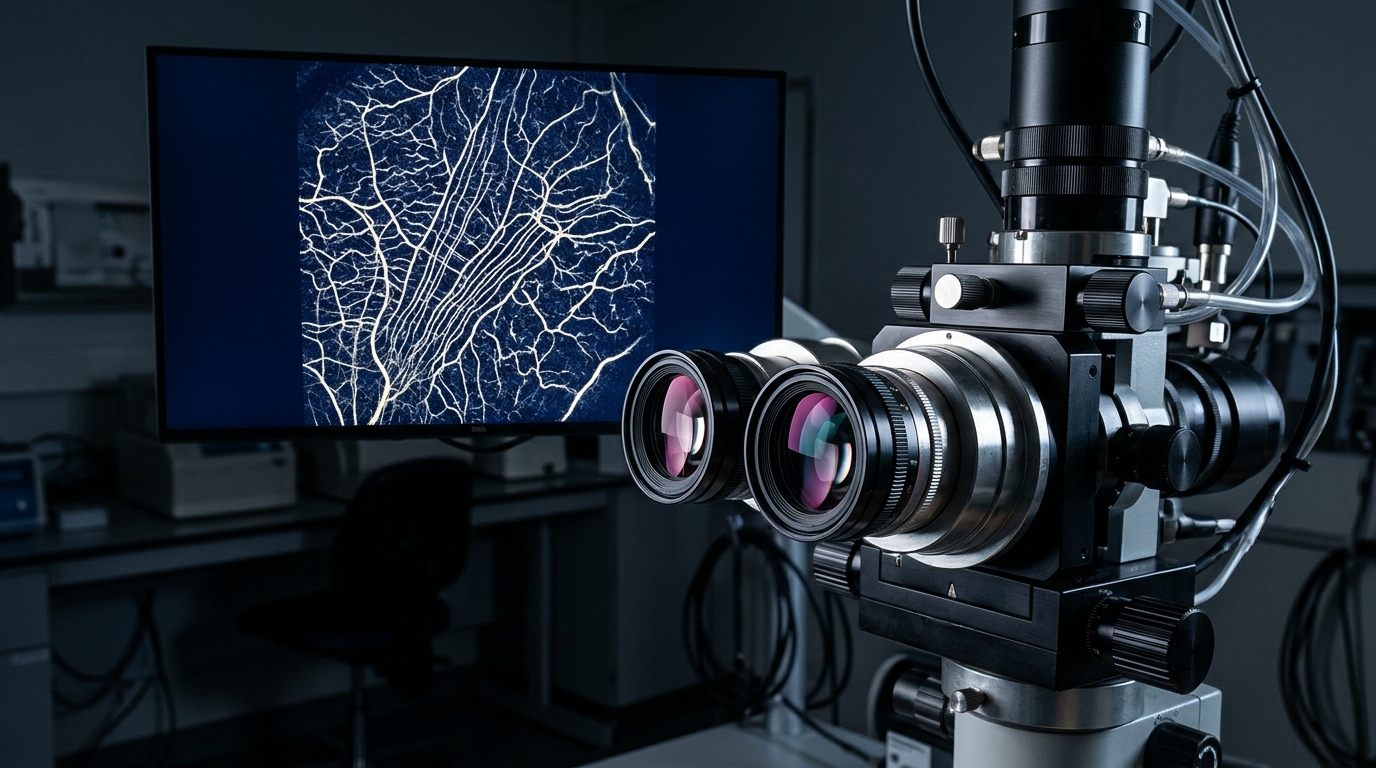
The cornea is the most densely innervated tissue in the body relative to its size, and the nerves visible under IVCM are small, unmyelinated C-fibres and thinly myelinated Aδ-fibres that originate from the trigeminal ganglion. Because these fibres share metabolic vulnerabilities with small-fibre nerves elsewhere in the body, the review’s authors argue that changes in their architecture may mirror peripheral nerve damage occurring elsewhere. IVCM allows clinicians to visualise these nerves at cellular resolution in a living patient, in real time, without a single incision, and automated analysis software has made it feasible to quantify nerve density and length across large cohorts.
The strongest evidence base by far lies in diabetic peripheral neuropathy, the most frequent long-term complication of diabetes. Across replicated cross-sectional and longitudinal studies, patients with diabetes show reductions in corneal nerve fibre density, branch density and fibre length, and these deficits appear before clinical symptoms and before abnormalities on conventional nerve conduction studies. A meta-analysis pooling 13 studies and 1,680 participants concluded that corneal nerve parameters were significantly reduced in patients with diabetes, especially when neuropathy was present. In a longitudinal assessment of 396 participants, corneal nerve fibre length was significantly lower in people with diabetic peripheral neuropathy than in diabetic patients without neuropathy, at 14.0 versus 19.1 mm/mm².
The predictive value of these measurements is a central part of the DPN story. A corneal nerve fibre length threshold below 14.9 mm/mm² predicted the development of diabetic peripheral neuropathy with a sensitivity of 0.82 and a specificity of 0.69 in one prospective cohort, and fibre length provided prognostic information up to six years before diagnosis, with an optimal cut-off of 14.1 mm/mm² yielding 67% sensitivity and 71% specificity. A sustained abnormal fibre length reading across at least half of a two-year observation period outperformed single measurements for predicting incident neuropathy, achieving an area under the curve of 0.763. Corneal nerve fibre density also correlated inversely with the Total Neuropathy Score, and fibre length in the inferior whorl region of the cornea emerged as the most sensitive early marker of diabetic sensorimotor polyneuropathy.
Artificial intelligence is now being layered onto this imaging approach. A convolutional neural network achieved an area under the curve of 0.83 for diagnosing diabetic peripheral neuropathy, outperforming a widely used automated analysis tool, while a ResNet-50 model with visualisation of its decision regions reached a sensitivity of 0.91, a specificity of 0.93 and an area under the curve of 0.95 across type 1 diabetes, type 2 diabetes and prediabetes. A vision transformer model pushed further, reporting an area under the curve of 0.99 with 98% sensitivity and 92% specificity without manual segmentation. A deep learning model combined with HbA1c achieved an area under the curve of 0.94 for predicting diabetic foot risk, and 0.93 when integrated with serum creatinine. The review cautions, however, that many of these AI studies lacked external validation datasets and may carry optimistic performance estimates.
In Parkinson’s disease, the picture is promising but less mature. Multiple studies have found reduced corneal nerve fibre density and length in patients compared with healthy controls, including in newly diagnosed patients whose skin biopsies showed normal epidermal nerve density, suggesting IVCM may detect small-fibre changes earlier than skin biopsy in this condition. Cross-sectional data show fibre density and length declining progressively from healthy individuals through prodromal to manifest Parkinson’s disease. Corneal nerve fibre loss has also been linked to cognitive impairment, with fibre density correlating positively with Montreal Cognitive Assessment scores, and to autonomic dysfunction, where corneal parameters distinguished patients with autonomic involvement from those without. In the single prospective study, patients in the lowest baseline quartile of fibre density experienced significantly greater motor deterioration over 12 months than those in the highest quartile. The review notes that applications such as prodromal detection, phenotyping and prognostication rest largely on small, single-centre studies.
For cognitive impairment and dementia, the review highlights comparisons between IVCM and established neuroimaging measures. In one cross-sectional study, the diagnostic accuracy of corneal nerve measures was comparable to medial temporal lobe atrophy scoring for dementia and superior to it for mild cognitive impairment, with higher sensitivity and specificity in the MCI group. In a prospective cohort of 107 people with mild cognitive impairment followed for a mean of 2.6 years, 33 progressed to dementia, and baseline corneal measures were lower in progressors, with reported prognostic accuracy of 72% to 75% for corneal confocal microscopy versus 68% to 69% for brain volumetry. Alzheimer’s disease patients also showed significantly lower fibre length, density and branch density, along with reduced corneal sensitivity, compared with healthy controls. The authors caution that this literature derives largely from overlapping single-centre cohorts, limiting independent validation.
Multiple sclerosis represents one of the stronger evidence bases after diabetic neuropathy. Two prospective cohorts with two-year follow-up demonstrated progressive corneal nerve loss in association with worsening disability: in relapsing-remitting MS, fibre area and fibre width decreased significantly as the Expanded Disability Status Scale increased, and changes in fibre density and length correlated with changes in disability scores. Cross-sectional studies have found reduced fibre density, length and fractal dimension across clinically isolated syndrome, relapsing-remitting and secondary progressive disease courses. However, the longitudinal cohorts remain small, and whether IVCM adds information beyond MRI or retinal nerve fibre layer assessment has not been formally established.
Elsewhere, the evidence thins considerably. In schizophrenia, a single cross-sectional study of 62 participants reported reduced fibre density, length and branch density, with fibre length achieving an area under the ROC curve of 88% for distinguishing patients from controls, exceeding the 73% achieved by cingulate gyrus volume, though neither measure correlated with symptom severity and confounders such as dry eye and antipsychotic exposure were not formally assessed. In amyotrophic lateral sclerosis, three small cross-sectional studies produced discordant results: two reported corneal nerve loss and correlations with bulbar severity, while one found no significant differences in standard nerve parameters. In migraine, two cross-sectional studies showed reduced fibre parameters relative to controls but no consistent relationship with headache frequency, duration or pain intensity. In chronic inflammatory demyelinating polyradiculoneuropathy, a single prospective cohort of 17 patients found that baseline immune-cell infiltrates above 30 cells/mm² predicted clinical progression over 18 months with 100% sensitivity and specificity, figures the authors describe as statistically fragile given the sample size. Evidence in tauopathies remains predominantly pre-clinical, drawn from transgenic mouse models showing corneal nerve degeneration that in some cases preceded memory deficits.
The review’s authors are clear that several hurdles stand between these findings and routine clinical use. Heterogeneity in confocal platforms, image acquisition protocols, image sampling strategies and analysis software can produce substantially different nerve measurements for the same tissue, with different platforms yielding sub-basal nerve density estimates that differ by two- to three-fold. Ageing, dry eye disease, diabetes without neurological involvement, contact lens wear and prior ocular surgery can all confound corneal nerve measurements. Direct evidence that IVCM provides incremental diagnostic or prognostic value over established biomarkers remains limited across most conditions. The authors call for standardised acquisition and analysis protocols, prospective multicentre longitudinal studies, appropriate adjustment for confounders and direct comparison with established biomarkers before broader clinical translation. For now, the cornea’s nerves offer researchers a non-invasive window on small-fibre neurodegeneration, one that is most convincing in diabetes, increasingly credible in multiple sclerosis, and still exploratory elsewhere.
Subject of Research: Corneal nerve status measured by in vivo confocal microscopy as a potential adjunctive biomarker for neurodegenerative and neurological diseases
Article Title: Corneal nerve status as a biomarker for neurodegenerative and neurological diseases: a narrative review
Article References: Li, J., Sim, A. E. H., Yu, M., Liu, C., Lee, I. X. Y., Anam, A., Koh, J. S., & Liu, Y.-C. (2026). Corneal nerve status as a biomarker for neurodegenerative and neurological diseases: a narrative review. British Journal of Biomedical Science, 83, Article 17067. https://doi.org/10.3389/bjbs.2026.17067
Image Credits: AI Generated
Keywords: corneal nerves, in vivo confocal microscopy, diabetic peripheral neuropathy, Parkinson's disease, multiple sclerosis, mild cognitive impairment, dementia, biomarker, small-fibre neuropathy, artificial intelligence, narrative review, neurodegeneration
News Source: Cassandra Pierce. (November 5, 2026). Corneal nerves under the microscope: a window on nerve damage in diabetes, Parkinson’s and beyond. Scienmag.