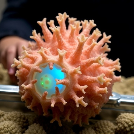
A coral-inspired scaffold that combines multi-walled carbon nanotubes with nano-hydroxyapatite has shown promise in restoring bone regeneration in steroid-induced osteonecrosis of the femoral head, a debilitating condition that can cause collapse of the hip joint. In a study published in Bone Research, researchers from Army Medical University and Sichuan University in China reported that the 3D-printed biomaterial did more than provide a physical framework for damaged bone. It also reshaped the local immune environment, helping inflammatory macrophages adopt a reparative identity. In rabbit models of steroid-induced osteonecrosis, this immune reprogramming was associated with increased blood-vessel formation, enhanced bone-forming activity and improved reconstruction of damaged femoral-head tissue.
Steroid-induced osteonecrosis of the femoral head, often abbreviated as SONFH, is one of the most serious complications associated with prolonged or high-dose glucocorticoid treatment. Reports suggest that it affects approximately 9% to 40% of patients exposed to these drugs, although the risk varies with dosage, treatment duration and individual susceptibility. Glucocorticoids can disrupt blood supply, lipid metabolism, bone-cell activity and immune regulation, gradually weakening the femoral head. As the disease progresses, the rounded upper end of the thigh bone may deform and collapse, ultimately requiring hip replacement. Core decompression, a procedure designed to reduce pressure and improve circulation inside the femoral head, is commonly used in early-stage disease, but it does not always restore the biological conditions required for lasting repair.
The researchers identified persistent inflammation as a major reason why damaged bone often fails to heal after surgical treatment. Macrophages, immune cells that remove debris and coordinate tissue responses, can adopt different functional states depending on signals in their surroundings. In simplified terms, M1-like macrophages promote inflammation, while M2-like macrophages support resolution of inflammation, tissue remodeling and repair. In SONFH, the investigators describe an “immune-freeze” microenvironment in which macrophages remain locked in a pro-inflammatory state instead of transitioning toward a reparative phenotype. This prolonged inflammatory activity can inhibit stem-cell function, impair the growth of new blood vessels and create conditions that are hostile to bone formation.
To address this problem, the team designed a scaffold inspired by the intricate, hierarchical pore network of coral. The structure was produced using three-dimensional printing and incorporated multi-walled carbon nanotubes, or MWCNTs, together with nano-hydroxyapatite, or nHA. MWCNTs are nanoscale carbon structures with distinctive mechanical, electrical and surface properties. In the scaffold, they were used primarily for their capacity to influence macrophage behavior and alter the cellular microenvironment. Nano-hydroxyapatite, meanwhile, resembles the mineral component of natural bone. Its inclusion was intended to create a biomimetic surface that could support cell attachment, proliferation and osteogenic differentiation, the process through which progenitor cells develop into bone-forming cells.
The resulting material was designed to combine two functions that are often treated separately in orthopedic biomaterials. Its porous architecture supplied physical space for tissue infiltration and helped reproduce aspects of the three-dimensional environment found in native bone. At the same time, its chemical and nanoscale composition provided signals capable of influencing immune cells. In laboratory experiments, the scaffold encouraged macrophages to move away from an inflammatory M1-like state and toward an M2-like reparative state. This change reduced the inflammatory character of the surrounding environment and generated signals more favorable to the activity of bone marrow mesenchymal stem cells. These multipotent cells are important participants in skeletal repair because they can migrate into damaged areas and contribute to the formation of new bone and supporting tissue.
The immunological effects were linked to activation of the PI3K-AKT signaling pathway, an intracellular communication system involved in cell survival, metabolism, proliferation and differentiation. When activated by extracellular cues, PI3K generates lipid-based signaling molecules that help recruit and activate AKT. The pathway can then influence transcriptional programs and other molecular processes governing cell behavior. In this study, the researchers associated PI3K-AKT activation with macrophage reprogramming and the broader regenerative response produced by the scaffold. The findings suggest that the material was not simply acting as an inert replacement for missing bone. Instead, it functioned as a local biological regulator, using the interaction between its surface and immune cells to influence the sequence of events required for tissue repair.
The consequences extended beyond macrophage behavior. The researchers observed that the altered immune environment promoted the migration of bone marrow mesenchymal stem cells and supported their osteogenic differentiation. The scaffold also enhanced angiogenesis, the formation of new blood vessels. This is a critical requirement for repairing osteonecrotic bone because regenerating tissue needs a supply of oxygen, nutrients and circulating cells. Blood vessels also provide important communication routes between the immune system and the skeletal system. By improving vascular development while reducing persistent inflammation, the scaffold addressed two closely connected barriers to recovery. In the rabbit model of SONFH, these effects were accompanied by more extensive new bone formation and improved reconstruction of defects compared with conventional scaffold treatments.
The study’s coral-inspired design reflects a growing shift in regenerative medicine toward materials that actively communicate with living tissue. Traditional orthopedic scaffolds are often evaluated mainly for their strength, porosity and ability to support cell growth. Those characteristics remain important, but they may not be sufficient when the injury is embedded in a chronically inflamed environment. A mechanically sound implant can fail to regenerate bone if immune cells continue releasing inflammatory signals that suppress vascularization and osteogenesis. The new findings support the idea that successful bone repair may require simultaneous control of structural, vascular and immunological processes. By combining a mineral phase that resembles bone with a nanotube component capable of influencing immune behavior, the researchers created a platform intended to coordinate these processes within the damaged femoral head.
The investigators emphasize that the work remains preclinical and that additional research will be needed before the scaffold can be considered for human treatment. Rabbit models can reproduce important features of osteonecrosis and provide valuable information about bone formation, vascularization and biomaterial compatibility, but they cannot fully predict how a complex implant will behave in patients. Future studies will need to examine long-term safety, degradation and retention of the scaffold, the fate of multi-walled carbon nanotubes, dose-dependent biological effects and the durability of the regenerated bone. Researchers will also need to determine whether the material can be manufactured consistently at clinical scale and whether its benefits remain effective in the presence of the diverse medical conditions and medication histories seen in people with SONFH. Even with those questions unresolved, the study offers a striking example of how immune reprogramming can be integrated into bone engineering. Rather than treating inflammation as a secondary complication, the coral-inspired scaffold targets it as a central driver of failed repair, opening a potentially powerful route toward therapies that restore both the structure of bone and the biological environment needed to rebuild it.
Subject of Research: Animals
Article Title: Coral-inspired immunoreprogramming scaffold reverses the “immune-freeze” microenvironment to promote bone regeneration in steroid-induced osteonecrosis of the femoral head
News Publication Date: 30 June 2026
Web References: https://doi.org/10.1038/s41413-026-00557-x; https://en.scu.edu.cn/
References: Bone Research; DOI: 10.1038/s41413-026-00557-x
Image Credits: Dr. Yan Xiong, Army Medical University (Third Military Medical University), China; Dr. Meng Tian and Dr. Pengde Kang, Sichuan University, China
Keywords: steroid-induced osteonecrosis of the femoral head; bone regeneration; coral-inspired scaffold; immunoreprogramming; macrophages; M1 macrophages; M2 macrophages; PI3K-AKT signaling; angiogenesis; osteogenesis; multi-walled carbon nanotubes; nano-hydroxyapatite; tissue engineering; regenerative medicine; biomaterials; mesenchymal stem cells
Tags: 3D-printed biomaterials for bone repairbiomaterial strategies for hip joint preservationcoral-inspired bone scaffoldenhancing angiogenesis in bone regenerationimmune environment reprogramming in bone healingimmune modulation in bone tissue engineeringmacrophage polarization for tissue regenerationmulti-walled carbon nanotubes in bone regenerationnano-hydroxyapatite for osteonecrosis treatmentregenerative approachesrole of immune cells in osteonecrosis recoverysteroid-induced osteonecrosis of the femoral head