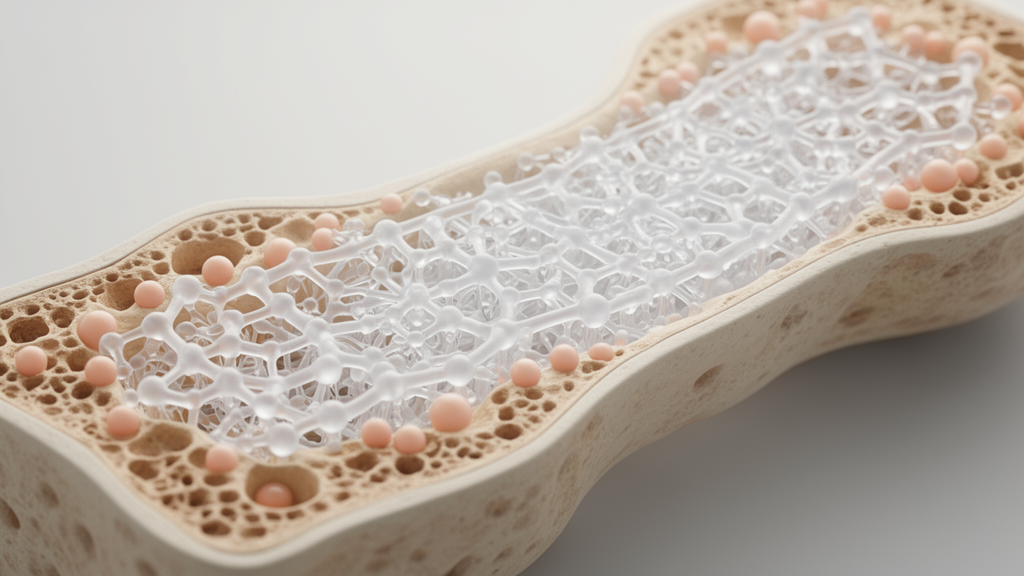
A comprehensive review of biodegradable biomaterials in bone tissue engineering has introduced a model-based clinical design framework aimed at synchronizing the degradation kinetics of temporary implants with the spatiotemporal stages of skeletal regeneration. Repairing critical-sized bone defects presents a persistent clinical challenge because spontaneous healing cannot occur when the physical gap exceeds the regenerative capacity of the host tissue. Writing in the Journal of Materials Science: Materials in Medicine, author Vahid Zarghami synthesized empirical observations, mechanistic formulas, and computational simulations across metals, ceramics, polymers, and composites. Rather than treating implant degradation as an immutable material property, the work outlines an engineering workflow that positions resorption rate as a programmable design parameter tailored to patient-specific biology, anatomical loading conditions, and defect geometry.
Natural bone healing progresses through a tightly regulated biological cascade spanning six consecutive phases, extending from initial mesenchymal condensation to mature structural remodeling. The biological timeline involves either intramembranous ossification, where bone forms directly without a cartilaginous precursor in flat bones such as the skull, or endochondral ossification, where repair proceeds through an intermediate cartilage template in long bones like the femur and tibia. In adult humans, intraosseous defects exceeding 2 to 2.5 centimeters generally fail to bridge spontaneously, establishing a critical threshold that demands temporary mechanical guidance and osteoconductive support. When an engineered scaffold degrades prematurely, the resulting mechanical instability causes structural failure; conversely, when degradation is excessively delayed, the persisting foreign material risks triggering chronic inflammation, fibrous tissue encapsulation, and mechanical stress shielding.
To navigate these biological constraints, the framework maps specific mathematical formulations to defect dimensions and anatomical scenarios. Small fracture gaps under one millimeter, which typically consolidate within six to eight weeks through direct intramembranous fusion under minimal strain gradients, are well described by linear mechano-regulatory algorithms. In contrast, intermediate defects nearing critical thresholds display sensitive endochondral repair sequences that correspond to sigmoidal critical volume models. These sigmoidal formulations capture the characteristic lag phase, rapid tissue accumulation, and eventual plateauing observed during callus stiffening and mineral deposition. For larger, supra-critical or segmental defects, predictive accuracy requires multi-physics formulations, including diffusion-reaction partial differential equations and continuum-level finite element simulations that track fluid velocity, shear strain, and cellular migration under physiological loading.
The physical mechanisms governing material breakdown vary fundamentally across biomaterial classes, necessitating distinct mathematical modeling paradigms for each group. Biodegradable metals such as magnesium, iron, and zinc alloys provide the high initial compressive strength exceeding 200 megapascals and fatigue resistance required for high-load anatomical sites like the femur and spine. However, pure magnesium exhibits a negative standard electrode potential of minus 2.37 volts versus the standard hydrogen electrode, causing rapid anodic dissolution, excessive hydrogen gas evolution, and local tissue alkalinization in chloride-rich physiological fluid. To manage this reactivity, researchers employ high-purity processing to eliminate cathodic impurities such as iron and nickel, while incorporating rare earth elements like yttrium, neodymium, or gadolinium to passivate surface oxide layers. Advanced formulations such as AZ31-GS leverage microalloying with gadolinium and scandium to establish core-shell intermetallic structures that substantially diminish Volta potential differences, lowering cathodic current density and in vitro degradation rates by more than half.
Iron and zinc biometals display contrasting electrochemical trajectories that define their respective computational requirements. Pure iron exhibits an electrode potential of minus 0.44 volts, generating a dense, passivating oxide film that degrades ten to twenty times slower than the rate of skeletal tissue formation, thereby raising risks of long-term mechanical stress protection and sustained inflammation. Accelerating iron breakdown requires engineered micro-galvanic couples, such as alloying with manganese or gold, alongside porous architectures that elevate the exposed surface area. Zinc occupies an intermediate kinetic regime with a potential of minus 0.76 volts, naturally degrading over six to twelve months without releasing gaseous byproducts, though it necessitates microalloying with calcium, magnesium, or strontium to achieve sufficient load-bearing strength. For metallic implants, the framework recommends mechanobiological finite element models that maintain local octahedral shear strains within an osteogenic threshold of two to five percent, ensuring dynamic stress transfer as the scaffold loses stiffness.
In contrast to metallic corrosion, biodegradable ceramics and aliphatic polymers degrade via surface dissolution and volumetric hydrolysis, requiring diffusion-reaction partial differential equation models. Workhorse calcium phosphate ceramics, including hydroxyapatite and beta-tricalcium phosphate, mimic bone mineral architecture and release osteogenic calcium and phosphate ions upon dissolution. While stoichiometric hydroxyapatite exhibits low solubility with a solubility product of approximately ten to the power of minus fifty-eight, beta-tricalcium phosphate has a higher solubility product near ten to the power of minus twenty-nine and undergoes active cellular resorption by osteoclasts. Designing biphasic calcium phosphate scaffolds with specific phase ratios allows tuned functional lifespans: a seventy-thirty hydroxyapatite to beta-tricalcium phosphate ratio resorbs over twelve to eighteen months, whereas an eighty-twenty ratio extends functional stability past eighteen to twenty-four months.
Aliphatic polyesters, including polyglycolic acid, polylactic acid, and polycaprolactone, resorb through volumetric ester bond scission into biocompatible monomers that enter native metabolic pathways such as the Krebs cycle. The hydrolytic degradation rate follows a defined material sequence: polyglycolic acid hydrolyzes fastest, followed by poly(lactic-co-glycolic acid), polylactic acid, and polycaprolactone, which can persist for two to three years. A primary operational hazard in polymer design is autocatalytic bulk erosion, in which fluid penetrates thick implants faster than ester bonds hydrolyze, trapping acidic degradation products in the interior core and accelerating premature mechanical disintegration. Mitigation approaches include incorporating basic ceramic fillers to buffer local acidity, optimizing copolymer ratios, and applying structural techniques such as stereochemical locking, where polylactic acid stereocomplexes form physical crosslinks within polyglycolic acid matrices to stabilize amorphous chains against water diffusion.
The review consolidates these engineering principles into an operational four-step clinical design methodology. Step one defines the biological target by calculating an estimated patient-specific healing timeline. Using a baseline rate derived from tibial fractures in healthy non-smoking adults, the conceptual formula scales healing duration by multiplying defect size in centimeters by five weeks per centimeter, and subsequently applying dimensionless patient and anatomical adjustment coefficients. Patient factors account for systemic biological conditions; for instance, type 2 diabetes reduces osteogenic and angiogenic potential, expanding the healing coefficient to between 1.4 and 1.6, while advanced age or smoking delays repair by thirty to fifty percent. Anatomical factors incorporate vascularization and regional loading differences, noting that mandibular defects heal approximately thirty percent faster than weight-bearing tibial sites, and metaphyseal regions repair ten to twenty percent faster than diaphyseal bone.
Steps two and three guide material selection and degradation tuning based on site-specific mechanical demands and the biological healing cascade. Step two directs high-load defects toward metallic alloys, moderate-load sites requiring fifty to two hundred megapascals toward ceramic-polymer composites, and non-load-bearing sites like craniofacial defects toward porous ceramics or biopolymers. Step three calibrates material kinetics across consecutive repair stages: inflammatory, soft callus, hard callus, and remodeling. Biodegradable metals are tuned via mechanical strain thresholds, bioceramics via dissolution rate constants matched to bone synthesis, and polymers via copolymer ratios designed to maintain structural stability through hard callus bridging before undergoing complete resorption during final remodeling.
Step four establishes a tiered experimental validation workflow culminating in a provisional quantitative metric termed the Synchronization Index. Testing begins with phase-specific in vitro immersion in media simulating diverse biological environments, such as inflammatory cytokines and low pH, followed by longitudinal in vivo micro-computed tomography and mechanical testing across key tissue transitions. The proposed Synchronization Index quantifies alignment by calculating the normalized area between the experimental implant mass loss curve and an idealized, patient-specific target bone formation curve on a dimensionless zero-to-one scale. In visual decision algorithms, an index value meeting the target threshold allows progress toward clinical translation, whereas substantial asynchrony redirects developers through an iterative redesign loop to adjust alloy composition, polymer crosslinking, or architectural porosity.
Despite outlining a structured design roadmap, the author emphasizes that the timeline formulas and the Synchronization Index remain conceptual tools that require rigorous preclinical validation. Because the linear heuristics cannot replace complex clinical judgement or address non-linear interactions among multiple patient comorbidities, extensive in vivo studies in large animal models are necessary to establish empirical thresholds. Translating the framework into routine orthopedic practice will require integrating patient-specific timeline formulas and multi-physics computational models into accessible clinical decision-support software, alongside targeted studies evaluating scaffold degradation within compromised biological environments such as elderly or diabetic populations.
Subject of Research: Model-driven framework for designing biodegradable biomaterials in bone tissue engineering
Article Title: Synchronizing degradation with regeneration: a model-driven framework for designing biodegradable biomaterials in bone tissue engineering
Article References: Zarghami, V. (2026). Synchronizing degradation with regeneration: a model-driven framework for designing biodegradable biomaterials in bone tissue engineering. Journal of Materials Science: Materials in Medicine, 37(1), Article 112. https://doi.org/10.1007/s10856-026-07102-6
Image Credits: AI Generated
DOI: 10.1007/s10856-026-07102-6
Keywords: biodegradable biomaterials, bone tissue engineering, critical-sized defects, degradation kinetics, mechanobiology, finite element modeling, diffusion-reaction equations, magnesium alloys, zinc alloys, iron biomaterials, biphasic calcium phosphate, polylactic-co-glycolic acid, autocatalytic erosion, synchronization index, scaffold design, bone regeneration
News Source: Ophelia Keating. (November 19, 2026). Computational Framework Matches Biodegradable Implant Resorption Rates to Bone Healing Timelines. Scienmag.