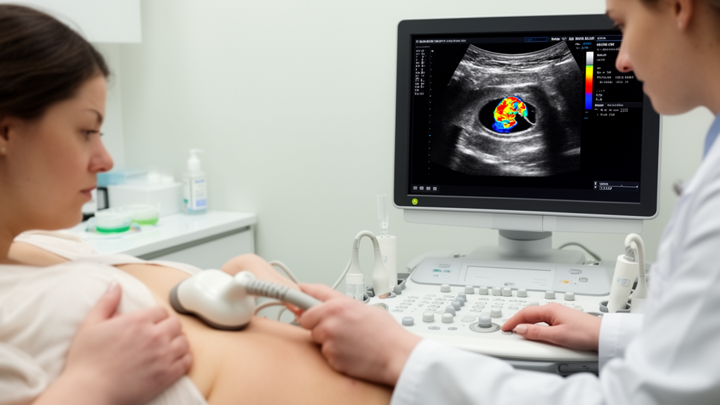
Combining automated breast ultrasound with conventional two-dimensional ultrasound improved the preoperative differentiation of benign and malignant intraductal breast lesions in a single-center study of 200 patients. The combined diagnostic model achieved an area under the receiver operating characteristic curve of 0.90, compared with 0.81 for two-dimensional ultrasound alone and 0.86 for automated breast ultrasound alone. Its reported sensitivity was 90.7% and specificity was 89.2%.
The study enrolled patients hospitalized with pathologically confirmed intraductal breast lesions at one hospital from April 2025 to April 2026. The cohort included 132 benign cases and 68 malignant cases. Every participant underwent both conventional two-dimensional ultrasound and automated breast ultrasound, or ABUS.
Investigators compared ultrasonographic features between the benign and malignant groups, then applied univariate and multivariate logistic regression to identify independent risk factors and construct a combined diagnostic model. Investigators used receiver operating characteristic curves to compare the diagnostic efficiency of the combined model with the two imaging approaches evaluated separately. Malignant lesions differed from benign lesions in several ultrasound features.
The malignant group had significantly higher incidences of irregular shape, microcalcification, abundant blood flow, duct dilatation of at least 5 mm, a convergence sign, lesion extension toward the nipple, and marginal spiculation or angulation. Each of these seven associations was statistically significant at P < 0.05, and multivariate logistic regression identified them as independent risk factors for malignancy. The model’s imaging variables therefore captured several distinct aspects of lesion appearance: irregular morphology, the presence of microcalcification, blood flow classified as Adler grade II–III, duct dilatation of at least 5 mm, convergence of tissue features, extension toward the nipple, and spiculation or angulation at the margin.
The analysis linked their combined presence with the likelihood that a pathologically confirmed lesion was malignant. The results indicate that ABUS and conventional two-dimensional ultrasound together produced favorable diagnostic performance for preoperative benign–malignant differentiation of intraductal breast lesions. In the studied population, the combined approach performed better than either examination alone according to the reported area under the curve.
The study therefore supports the combined use of the two ultrasound methods as an imaging aid for this specific group of lesions, rather than establishing a general benefit across all types of breast abnormalities. Several features of the study limit how broadly the findings can be interpreted. It was conducted in a single hospital and included 200 hospitalized patients with pathologically confirmed intraductal breast lesions, including 68 malignant cases.
The model was developed and evaluated in that same single-center cohort, and the supplied results do not establish performance in other hospitals, screening populations, or patients with a wider range of breast findings. Because the study was limited to one institution and a relatively small malignant subgroup, the combined model’s diagnostic performance should be viewed as specific to the evaluated cohort. The reported AUC, sensitivity, and specificity quantify performance in this sample; they do not by themselves demonstrate that the model will maintain the same results in external populations.
Further evidence would be needed to determine how consistently the approach performs across different clinical settings. Overall, the study found that combining automated and conventional ultrasound produced the strongest reported discrimination between benign and malignant intraductal breast lesions. The analysis linked improved classification to seven ultrasound characteristics identified as independent risk factors in the cohort.
Its findings offer an imaging-based model for preoperative assessment in hospitalized patients with pathologically confirmed intraductal lesions, while the single-center design defines the boundary of the evidence.
Subject of Research: Preoperative ultrasound diagnosis of intraductal breast lesions
Article Title: Diagnostic value of combined automated breast ultrasound system and conventional two-dimensional ultrasound for intraductal breast lesions
Article References: Yang, Q., Yi, R., Guo, F., Gou, T., Cai, L., Wang, S., Zhang, Y., & Luo, Y. (2026). Diagnostic value of combined automated breast ultrasound system and conventional two-dimensional ultrasound for intraductal breast lesions. BMC Medical Imaging. https://doi.org/10.1186/s12880-026-02832-z
Image Credits: AI Generated
DOI: 10.1186/s12880-026-02832-z
Keywords: ABUS, breast ultrasound, intraductal lesions, breast cancer, diagnostic imaging, two-dimensional ultrasound, microcalcification, duct dilatation, logistic regression, diagnostic accuracy, Diagnostic, value
News Source: Ophelia Keating. (October 20, 2026). Combined Ultrasound Improves Intraductal Breast Lesion Diagnosis. Scienmag.