


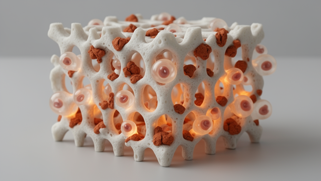
Engineers in India have 3D printed porous bone scaffolds from hydroxyapatite reinforced with tiny amounts of halloysite nanoclay, and their laboratory tests show that a small dose of the clay makes the scaffolds measurably harder and more welcoming to bone-derived cells than the ceramic alone. The work, published in Frontiers in Bioengineering and Biotechnology, was carried out by A. J. Ashwin, J. M. Jafferson and Davidson Jebaseelan at Vellore Institute of Technology, Chennai, and describes a complete fabrication-and-testing pipeline for a composite aimed at hard tissue repair.
Hydroxyapatite is a calcium phosphate mineral that closely resembles the inorganic component of natural bone, which is why it has long been treated as a benchmark material for bone reconstruction. It is biocompatible and bioactive, but it is also brittle, a weakness that has kept pure hydroxyapatite scaffolds out of load-bearing roles. The Chennai team’s strategy was to reinforce the ceramic with halloysite nanoclay, a naturally occurring tubular clay mineral that is considered biocompatible and that brings a rigid, high-aspect-ratio geometry well suited to carrying mechanical load inside a ceramic matrix.
The scaffolds were made by robocasting, a direct-ink-writing form of additive manufacturing in which a dense ceramic paste is extruded layer by layer through a fine nozzle under computer control. The researchers designed a 10 by 10 by 7 millimetre scaffold, sliced it at 50 percent infill, and printed it with a 0.8 millimetre nozzle. Getting the paste right was a study in itself. Hydroxypropyl methylcellulose served as the binder, and three concentrations were tested. At 1 weight percent the paste was too runny, dripping and sagging under its own weight; at 2 weight percent it was too stiff, curling and snapping during extrusion. The 1.5 weight percent paste showed balanced flow, forming straight, uniform strands with good spanning ability between printed pillars.
Rheological measurements backed up what the extrusion trials showed. All three pastes behaved as non-Newtonian shear-thinning fluids, with viscosity starting near 6,000 pascal-seconds at low shear rates and dropping as shear increased. The 1.5 percent paste developed the highest initial yield stress, spiking above 400 pascals, indicating a sturdy, entangled polymer-ceramic network at rest that lubricated smoothly once flowing. The weaker pastes peaked around 200 pascals. The chosen formulation behaved as a Bingham plastic, holding its shape after deposition without sagging or necking.
After printing, the green scaffolds were dried at 120 degrees Celsius for 12 hours and sintered at 1,100 degrees Celsius with a four-hour soak, a temperature chosen to stay below the roughly 1,200 degree threshold where hydroxyapatite can decompose into secondary calcium phosphates, while still hot enough to drive solid neck formation between ceramic particles. Field-emission electron microscopy confirmed that the nanoclay survived sintering and remained dispersed in the matrix. Energy-dispersive X-ray spectroscopy detected aluminium and silicon, the chemical fingerprints of halloysite, in every reinforced scaffold, and micropores between 0.1 and 6 micrometres along with nanopores between 2 nanometres and 1 micrometre were found throughout.
Dispersion proved to be the deciding variable. In scaffolds with 1 and 2 weight percent nanoclay the reinforcement was distributed homogeneously, but at 3 weight percent the team observed microcracks, clustering and clumping, caused by stress from agglomerated excess reinforcement. That observation capped the useful loading range. Mechanical testing told a consistent story. Vickers microhardness rose from 245.67 HV for pure hydroxyapatite to 257.32 HV at 1 percent clay and 271.67 HV at 2 percent, then essentially plateaued at 274.67 HV at 3 percent, where diagonal edge breakage during testing reflected the non-homogeneous mixing. The researchers attribute the gains to efficient load transfer from the weaker ceramic phase to the stronger nanotubes, which bridge microcracks and deflect crack propagation. Porosity, measured by liquid displacement using Archimedes’ principle, stayed in a narrow band, descending from 39.18 percent for pure hydroxyapatite to 38.21 percent at 3 percent clay.
Biological performance also improved with clay loading. In MTT viability assays on MG-63 osteoblast-like cells, pure hydroxyapatite showed viability declining from 100 percent to roughly 71 to 80 percent at the highest concentrations. With 1 percent nanoclay, viability stayed between about 87 and 95 percent at low and mid doses and around 74 percent at the highest dose. At 2 percent clay, viability was around or slightly above 100 percent at low concentrations and remained in the 80 to 90 percent range even at the highest dose, and at 3 percent it stayed close to or above 100 percent across most concentrations. All formulations remained above the roughly 70 percent threshold generally regarded as non-cytotoxic in vitro.
The Alamar Blue proliferation assay reinforced the same trend. Pure hydroxyapatite fell from 91.63 percent viability at 75 micrograms per millilitre to 79.69 percent at 600 micrograms per millilitre, while the 1 percent composite ranged from 92.42 to 82.43 percent and the 2 percent composite from 97.82 to 84.74 percent. The 3 percent composite performed best, peaking at 100.34 percent at the lowest concentration. The authors attribute the boost to biologically active silicon ions released from the clay, along with increased nanoscale surface roughness and hydrophilicity that favour protein adsorption and cell attachment. Swelling tests in phosphate-buffered saline showed modest fluid uptake, rising quickly in the first 12 hours and reaching a stable plateau by 48 hours, with the reinforced scaffolds absorbing slightly more fluid than pure hydroxyapatite but without the volumetric expansion typical of polymeric hydrogels.
Antimicrobial testing against Staphylococcus aureus by the Kirby-Bauer disk diffusion method produced more modest results. Ciprofloxacin, the positive control, produced a 25.33 millimetre zone of inhibition. Pure hydroxyapatite at 500 micrograms produced an 8.66 millimetre zone, while the composites produced zones of 4 to 6 millimetres depending on loading. The researchers interpret these smaller zones as evidence of intrinsic, contact-mediated antibacterial activity arising from surface interactions, electrostatic effects and nanotopographical stresses rather than the DNA-targeting mechanism of an antibiotic, and they argue that such localized, contact-active behaviour could help prevent early biofilm formation without the selective pressure that drives antibiotic resistance.
Overall, the study identifies the 2 weight percent nanoclay formulation as the best all-round composition, combining near-peak hardness, high cell viability and proliferation, and tolerable antibacterial behaviour at roughly 38 percent porosity. The authors suggest this composition is suitable for repairing cancellous bone, the spongy interior bone tissue that carries lower loads than cortical bone. It is worth emphasizing what these results do and do not show: all mechanical and biological findings come from in vitro laboratory tests on a single cell line and disk-diffusion assays, and the scaffolds have not yet been tested in animal models or in patients. The work does, however, map out a reproducible printing recipe and a clear dispersion limit for clay-reinforced hydroxyapatite, giving bone tissue engineering researchers a concrete starting point for the next stage of evaluation.
Subject of Research: Robocasted hydroxyapatite scaffolds reinforced with halloysite nanoclay for bone tissue engineering
Article Title: Advanced fabrication and characterization of robocasted halloysite-hydroxyapatite nanocomposite scaffolds for hard tissue regeneration
Article References: Ashwin, A. J., Jafferson, J. M., & Jebaseelan, D. (2026). Advanced fabrication and characterization of robocasted halloysite-hydroxyapatite nanocomposite scaffolds for hard tissue regeneration. Frontiers in Bioengineering and Biotechnology, 14, Article 1909411. https://doi.org/10.3389/fbioe.2026.1909411
Image Credits: AI Generated
DOI: 10.3389/fbioe.2026.1909411
Keywords: hydroxyapatite, halloysite nanoclay, robocasting, 3D printing, bone scaffolds, MG-63 cells, Vickers hardness, antimicrobial assay, sintering, tissue engineering, porosity, HPMC binder
News Source: Gregory Coleman. (October 28, 2026). Clay-Boosted 3D Printed Bone Scaffolds Show Stronger, More Cell-Friendly Results. Scienmag.



