Researchers in South Korea have built a microfluidic chip that separates and detects SARS-CoV-2 immunocomplexes in less than 30 seconds, offering a rapid alternative to both laboratory molecular testing and commercial lateral-flow antigen kits. The platform, developed by Gayathri Sureshkumar, To Thanh Truc Truong, Trung Nghia Tang and Tae Seok Seo and published in Advances in Industrial and Engineering Chemistry, performs an on-chip immunoassay on a disposable polymethyl methacrylate (PMMA) substrate filled with a 5.5% linear polyacrylamide (LPA) sieving matrix. Rather than relying on the dual-antibody, gold-nanoparticle mechanism of lateral-flow strips, the assay requires only a single fluorescently labeled antibody, and a positive or negative result is read directly from the presence or absence of an immunocomplex peak in the electropherogram. The team reports a detection limit of 5 µg/mL for the viral nucleocapsid protein, consistent performance across the alpha, beta and delta variants, and reliable detection in nine clinical oropharyngeal swab specimens confirmed beforehand by RT-qPCR.
The motivation stems from a persistent tradeoff in diagnostic design. Reverse-transcription polymerase chain reaction remains the gold standard for sensitivity and low viral-load detection, but it depends on laboratory infrastructure, trained personnel and multi-hour thermal cycling, which makes it poorly suited to decentralized outbreak response. Antigen-based rapid tests deliver results in minutes and enable mass screening outside laboratories, but at lower analytical performance. Microcapillary electrophoresis (µCE) has repeatedly proven itself as a point-of-care compromise, translating the resolving power of benchtop capillary electrophoresis onto a chip while cutting sample volume, reagent use and run time by orders of magnitude. Most µCE diagnostic work to date, however, targets nucleic acids, where mobility-based separation cleanly discriminates amplification products by fragment length. Prior handheld analyzers have resolved SARS-CoV-2 genetic markers to a smartphone and even identified 28 respiratory pathogens in a single multiplexed run, but in both cases detection depended on a prior PCR amplification step that adds hours, extra reagents and precise temperature control to the workflow.
Targeting proteins instead of nucleic acids removes the amplification bottleneck directly. Viral antigens and host antibodies exist at detectable levels in their native state, retaining their folded conformations and intact binding epitopes directly in patient swab lysates without denaturing chemical pre-treatment or thermal unfolding. Sample preparation is reduced to lysis, releasing the target protein into solution with no further processing. Yet miniaturizing protein capillary electrophoresis has lagged far behind its nucleic acid counterpart for several reasons. Proteins adsorb onto microchannel walls through electrostatic and hydrophobic forces, and at the microscale, where the surface-area-to-volume ratio is much higher than in standard fused-silica capillaries, this adsorption distorts electroosmotic flow, broadens bands, produces asymmetric peaks and ruins the reproducibility of migration times. Existing polymer coatings designed to suppress adsorption were developed on glass or fused-silica substrates, leaving their application to PMMA, a cheaper and more accessible substrate for disposable chips, largely unexplored.
Protein charge behavior poses a second challenge. Unlike DNA, which carries a constant negative charge from its phosphate backbone regardless of sequence, each protein has a distinct isoelectric point set by its acidic and basic residues, so its net charge and electrophoretic mobility depend on where the buffer pH sits relative to that value. Antigen-antibody complexes can have isoelectric points close to their unbound components, giving protein mixtures a narrower and less predictable range of charge-to-mass ratios than nucleic acid fragments of different lengths and making clean separation of bound from unbound species sensitive to buffer conditions. Surfactants add a third complication: sodium dodecyl sulfate, the workhorse of conventional protein electrophoresis, denatures proteins and can dissociate antigen-antibody complexes, making it unsuitable for binding-based immunoassays. The team selected Tris-borate-EDTA buffer at reduced ionic strength to maintain a stable alkaline pH, minimize Joule heating during high-field separation and leverage EDTA as a metal-ion scavenger to suppress metal-catalyzed protein aggregation and fluorophore quenching.
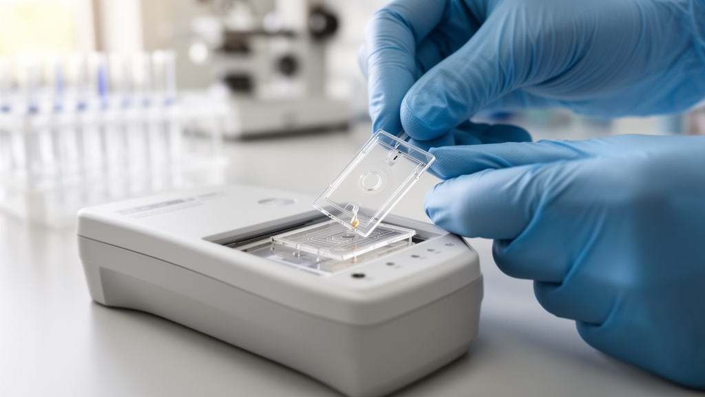
The chip itself is a three-layer laminate: a 3 mm-thick PMMA substrate patterned by CNC ball-end milling with a double-T microchannel 200 µm wide and 500 µm deep, sandwiched between pressure-sensitive adhesive films that seal the trenches. The layout includes a 6 mm-long injection channel connecting the sample and waste reservoirs and a 14.5 mm-long separation channel running from the stacking intersection to the anode reservoir, a path length sufficient to resolve protein complexes without excessive voltages. A 0.5 mm sample-stacking region in the injection arm accumulates the immunocomplex mixture before separation, boosting detection sensitivity. To suppress nonspecific protein adsorption, the channel surface was covalently functionalized through sequential oxygen plasma oxidation, EDC/NHS activation and grafting of amino-polyethylene glycol, replacing the native hydrophobic PMMA character with a hydrophilic, protein-repellent layer terminating in hydroxyl groups. Comparison between coated and uncoated channels showed a 78% reduction in residual albumin fluorescence, confirming that the passivation strategy enables adsorption-free migration and consistent band shape across replicate runs.
Operation proceeds in four stages. During electrokinetic injection, a field of 180 V applied at the waste reservoir with the sample reservoir held at 0 V drives the fluorescent mixture into the stacking region in roughly 10 seconds. Once fluorescence imaging confirms effective preconcentration at the stacking intersection, the electrode configuration switches to 320 V at the anode and 0 V at the cathode, launching the stacked sample into the main separation channel. The analytes then resolve into distinct bands according to their charge-to-size ratios and are detected within 30 seconds in a monitoring zone positioned near the anode reservoir. A custom dual-voltage circuit powered by a 12 V DC supply, with two DC-DC boost converters and a relay switch, alternates between low-field preconcentration and high-field separation through the same electrode set without reconfiguration. Fluorescence signals are acquired by an inverted microscope under 488 nm LED excitation, and a custom Python pipeline converts recorded video into electropherograms, extracting time-dependent intensity along a fixed detection line at 30 frames per second, with normalization and moving-average smoothing performed using open-source libraries.
Before testing viral targets, the team validated antibody migration against abundant background proteins. Albumin, at 66 kDa, migrated as a single sharp peak at 10 seconds, while fibrinogen, at 340 kDa, migrated substantially later at 22 seconds, consistent with its larger size and lower mobility. The purified Alexa Fluor 488-conjugated anti-nucleocapsid antibody alone produced a single sharp peak at 14 seconds, positioned between the two reference proteins as expected for its intermediate 150 kDa molecular weight. When albumin and fibrinogen were mixed at a 1:1 ratio, both peaks resolved cleanly at their individually determined migration times, and when the antibody was added at a 2:1:1 ratio, all three peaks again resolved at their expected positions without shift or interference, confirming that the antibody does not react with these background components. The authors note that nonspecific IgG, which is similar in size to the target antibody, would be expected to migrate close to it and could present a peak-overlap challenge that requires further investigation with a longer channel or higher voltage.
With peak identity established, the binding reaction was analyzed across a dilution series from 50 to 2.5 µg/mL of recombinant nucleocapsid protein. The faster-migrating peak corresponds to unbound antibody, which moves ahead due to its smaller size, while the slower peak corresponds to the antibody-nucleocapsid immunocomplex, which migrates more slowly due to its larger size and the altered charge density contributed by the antigen. At 50 µg/mL the two peaks eluted at 15 and 22 seconds, and the complex peak intensity decreased gradually with decreasing antigen concentration until it could no longer be resolved below 5 µg/mL, defining the detection limit. Absolute migration times shifted modestly between runs due to small variations in field strength, buffer conductivity or electroosmotic flow, but the relative migration time, the ratio of complex to antibody migration time, remained close to 1.5 throughout. Plotting complex peak height against concentration produced a nonlinear calibration curve with an R² of 0.9935. The authors attribute the detection limit to the antibody’s stoichiometric binding capacity, calculated at approximately 41.6 µg/mL of antigen, rather than to the resolving power of the separation itself, since the assay relies on direct fluorescence with no enzymatic signal amplification.
Clinical evaluation and specificity testing followed. Nine anonymized oropharyngeal swab samples obtained from Kyung Hee University Hospital at Gangdong were analyzed: six SARS-CoV-2 positive samples and three containing other common respiratory pathogens, respiratory syncytial virus A, adenovirus and influenza H1N1, each confirmed by RT-qPCR. Clinical specimens were lysed for one hour in a buffer of phosphate-buffered saline, Triton X-100 and potassium chloride, then mixed with the purified antibody before loading. All six positive samples resolved two distinct peaks corresponding to free antibody and the immunocomplex, confirming detection of the nucleocapsid protein directly from clinical specimens without nucleic acid amplification. The three non-SARS-CoV-2 samples produced only a single unbound-antibody peak, indicating negligible cross-reactivity with proteins from the other pathogens. Absolute migration times shifted slightly relative to buffer-only mixtures, likely reflecting ionic strength or residual matrix components, but resolution was preserved in every sample. Recombinant antigens corresponding to the alpha, beta and delta variants were likewise resolved into well-defined peaks within 30 seconds, demonstrating consistent performance regardless of strain variation.
The authors acknowledge current limitations: the assay provides only semi-quantitative detection over a narrow concentration range, because the complex signal saturates at higher antigen concentrations and the sample matrix influences migration and signal, and separating proteins with similar sizes remains difficult under the current conditions. Even so, the platform’s sub-minute separation time compares favorably with the roughly 15 minutes required by commercial rapid antigen tests, and its compatibility with automation positions it as a scalable route toward decentralized point-of-care testing. Future work will focus on improving quantification, enhancing separation resolution and integrating the µCE chip into a portable point-of-care system. Beyond SARS-CoV-2, the researchers argue that the mobility-based readout establishes protein-based microcapillary electrophoresis as a practical route to rapid antigen testing that could be extended to other protein biomarkers of infectious disease.
Subject of Research: Development of an on-chip microcapillary electrophoresis platform for rapid, amplification-free detection of SARS-CoV-2 nucleocapsid immunocomplexes in clinical samples
Article Title: Rapid and sensitive on-chip microcapillary electrophoresis for discrete SARS-CoV-2 immunocomplex diagnostics
Article References: Sureshkumar, G., Truong, T. T. T., Tang, T. N., & Seo, T. S. (2026). Rapid and sensitive on-chip microcapillary electrophoresis for discrete SARS-CoV-2 immunocomplex diagnostics. Advances in Industrial and Engineering Chemistry, 2(1), Article 14. https://doi.org/10.1007/s44405-026-00054-z
Image Credits: AI Generated
DOI: 10.1007/s44405-026-00054-z
Keywords: microcapillary electrophoresis, SARS-CoV-2, nucleocapsid protein, immunocomplex, point-of-care diagnostics, PMMA microfluidics, antigen detection, linear polyacrylamide, fluorescence detection, clinical swabs, Rapid, sensitive
News Source: Kristina Jarvis. (October 26, 2026). Chip-Based Electrophoresis Spots SARS-CoV-2 Antigen in Under 30 Seconds. Scienmag.