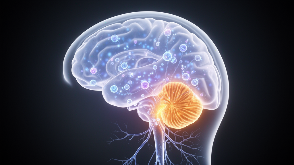
For decades, the cerebellum was dismissed as the brain’s humble movement coordinator, a structure that fine-tuned gait and balance while the cerebral cortex handled everything that makes us human. That view has collapsed. A new study from Stanford University School of Medicine, published in the Journal of Autism and Developmental Disorders, now provides some of the most detailed in vivo evidence yet that two folds at the back of the cerebellum, known as Crus I and Crus II, show measurable structural and neurochemical differences in autistic children, and that these differences track with the severity of one of autism’s most challenging features: restricted and repetitive behaviors.
The research team, led by John P. Hegarty II of the Department of Psychiatry and Behavioral Sciences, recruited 62 participants and ultimately obtained high-quality structural MRI data from 13 autistic children, 16 neurotypical control children, and a smaller exploratory group of seven autistic adolescents. Crucially, the pediatric sample was designed to be representative of the spectrum rather than a filtered subset. It included children with profound autism, intellectual disability, and significant language delays, populations that are routinely excluded from neuroimaging research and whose absence has long been suspected of masking biologically meaningful subgroups.
Scanning young autistic children without sedation is a formidable technical challenge, and the team relied on validated sleep-scanning protocols for children roughly six and under, with sufficient head and neck padding to minimize motion. Older children and adolescents were scanned awake after behavioral training on an MRI simulator, during which a head-mounted motion sensor provided real-time feedback paired with access to a preferred video until participants could hold still within two millimeters for at least five minutes. Similar proportions of autistic and control children completed sleep versus awake scans, reducing the risk that arousal state confounded group comparisons.
To segment the cerebellar folds with precision, the researchers used DeepCERES, a newly developed artificial intelligence-based tool from the volBrain platform that delineates individual cerebellar lobules from ultra-high-resolution multimodal MRI. Trained raters visually inspected every scan before and after segmentation to confirm accurate separation of cerebellar tissue from surrounding cerebrospinal fluid. The results were striking: Crus I and Crus II were significantly thinner in autistic children than in neurotypical controls, with the effect most pronounced in the left hemisphere of Crus I and the right hemisphere of Crus II. Autistic children also showed reduced asymmetry in Crus II between hemispheres and marginally thinner left Crus I cortex compared with autistic adolescents, hinting at developmental trajectories that shift with age.
Structure was only half of the story. The study’s most technically ambitious component was the use of a novel proton magnetic resonance spectroscopy sequence, called MEGA-SPECIAL, to measure the brain’s principal excitatory and inhibitory neurotransmitters, glutamate and GABA, directly within the cerebellar hemispheres. The standard clinical sequence, MEGA-PRESS, suffers from a well-known weakness: co-edited macromolecule signals can contaminate GABA quantification by 40 to 60 percent. MEGA-SPECIAL employs more selective editing pulses, set at 1.9 and 7.5 parts per million, to suppress these macromolecule signals and improve localization. Single-voxel data were acquired from two-by-four-by-two-centimeter regions in the left and right cerebellar hemispheres over ten-minute acquisitions, with glutamate quantified using LCModel and GABA measured from the edited 3 ppm peak.
Quality control was rigorous. Spectra were included only if the full-width at half-maximum line width remained below 15 hertz, Cramer-Rao lower bounds stayed under 20 percent, and visual inspection confirmed reliable model fitting. Even so, spectroscopy proved harder to obtain than anatomy: of 42 children who attempted imaging, high-quality neurometabolite data were available from only 10 autistic children, nine neurotypical controls, and four autistic adolescents. Within that reduced subset, the researchers found no significant group differences in glutamate or GABA concentrations, a null result they interpret cautiously given the small numbers.
The correlations, however, were compelling. Across all participants regardless of diagnosis, thinner Crus I grey matter and smaller Crus II volume were associated with a higher ratio of excitatory glutamate to inhibitory GABA, an index of the excitation-inhibition balance that many theorists place at the heart of autism neurophysiology. The strongest relationships appeared in left Crus I, with a correlation of negative 0.70, and right Crus II, at negative 0.61, both surviving correction for multiple comparisons. Glutamate drove most of the effect, correlating at negative 0.78 with left Crus I thickness. In other words, the smaller the cerebellar fold, the more its chemistry tilted toward excitation.
When the team linked these brain measures to behavior, one domain dominated: restricted and repetitive behaviors, or RRB. Crus I volume correlated with self-injurious behavior in both hemispheres, reaching a correlation of 0.53 on the left that survived statistical correction. Within the autistic sample alone, the associations strengthened, with left Crus I volume correlating at 0.71 with self-injurious behavior. Neurochemistry told a parallel story. Right-hemisphere GABA levels tracked with self-injurious and restricted behaviors, and the single strongest spectroscopy finding, a correlation of 0.81 between glutamate and compulsive behaviors, survived correction for multiple comparisons. By contrast, relationships with social communication and sensory processing were modest, a pattern the authors attribute partly to the limited sensitivity of standard screening instruments to the social subdomains most influenced by cerebro-cerebellar circuitry.
These findings converge with a substantial body of post-mortem and translational work. Autopsy studies have repeatedly documented reduced Purkinje cell density, diminished dendritic arborization, and altered cellular size and shape in the posterolateral cerebellar hemispheres, some of the most consistent neuropathological findings in autism. Mouse models reinforce the circuit-level logic: genetic knockouts of autism-associated genes such as SHANK2 and PTEN produce Purkinje cell loss, reduced neuroplasticity, and the emergence of social and repetitive behavioral traits. The cerebellum also develops earlier than the cortex, with explosive growth in the late second and third trimesters, a window in which cerebellar hemorrhage in premature infants is associated with markedly elevated rates of autism, language delay, and intellectual disability. The authors propose that early cellular alterations disrupt cerebellar modulation of cortical development, derailing the feedback loops that optimize neural circuit dynamics for social, cognitive, and behavioral skill acquisition.
The study is explicitly preliminary, and the authors are candid about its limits. The spectroscopy samples are small, the adolescent group lacked age-matched neurotypical controls, and neurometabolite concentrations are an indirect proxy for excitation-inhibition neurophysiology rather than a direct measure of it. Psychoactive medication use was not specifically excluded, and the novel MRS sequence, while suppressing macromolecule contamination, still requires cross-validation against standard sequences by independent laboratories. Yet the implications are tantalizing. If a cerebellar phenotype, defined by structure and excitation-inhibition balance, characterizes a identifiable subgroup of autistic children, non-invasive biomarkers could eventually guide patient stratification and even inform neuromodulation therapies targeting cerebro-cerebellar circuits. For a condition affecting roughly one in 31 children in the United States and still lacking any biological markers for clinical care, the idea that answers might lie in a structure long dismissed as the brain’s afterthought is precisely the kind of paradigm shift the field has been waiting for.
Subject of Research: Cerebellar structure and excitation-inhibition balance in autism spectrum disorder
Article Title: Cerebellar Crus I and Crus II Structure and Excitation/Inhibition Balance in Autism Spectrum Disorder
Article References: Hegarty, J. P., II, Komaki, H., Gu, M., Young, K., Ogbonnaya, N., Spielman, D. M., & Hardan, A. Y. (2026). Cerebellar Crus I and Crus II Structure and Excitation/Inhibition Balance in Autism Spectrum Disorder. Journal of Autism and Developmental Disorders. https://doi.org/10.1007/s10803-026-07529-4
Image Credits: AI Generated
DOI: 10.1007/s10803-026-07529-4
Keywords: autism spectrum disorder, cerebellum, Crus I, Crus II, glutamate, GABA, excitation-inhibition balance, MR spectroscopy, cortical thickness, restricted and repetitive behaviors, neurodevelopment, MEGA-SPECIAL
News Source: Cassandra Pierce. (October 8, 2026). Cerebellar Structure and Brain Chemistry Imbalance Linked to Autism Traits in Children. Scienmag.