Every year, millions of people survive a heart attack thanks to rapid reopening of the blocked coronary artery, yet a troubling paradox follows: restoring blood flow itself inflicts additional damage on the starving heart muscle. This phenomenon, known as myocardial ischemia-reperfusion injury or MIRI, remains one of cardiology’s most stubborn unsolved problems. Now a comprehensive review published in the Journal of Translational Medicine by Xue Guan, Luqiao Wang and colleagues at the First Affiliated Hospital of Kunming Medical University argues that the answer may lie in a structure most people have never heard of: the mitochondria-associated endoplasmic reticulum membrane, or MAM. These tiny bridges between two of the cell’s most important organelles, the authors contend, act as a master control platform that links fat metabolism, calcium signaling and a form of iron-driven cell death called ferroptosis into a single destructive cascade during reperfusion.
MAMs are not organs in the conventional sense but specialized zones of close physical contact between the endoplasmic reticulum, the cell’s protein and lipid factory, and mitochondria, its power plants. The two membranes are held roughly 10 to 25 nanometers apart by protein tethers, including the mitofusin 2 bridge, the voltage-dependent anion channel VDAC1 on the mitochondrial outer membrane paired with the inositol trisphosphate receptor IP3R on the ER via the chaperone GRP75, and additional connectors such as PTPIP51, VAPB and ATAD3A. This architecture is far more than scaffolding. At these contact points the cell exchanges calcium from ER stores into mitochondria, synthesizes and shuttles phospholipids, coordinates mitochondrial fission through the dynamin-related protein Drp1, and monitors mitochondrial quality through mitophagy pathways involving PINK1 and Parkin. In cardiomyocytes, which beat billions of times over a lifetime and depend on relentless ATP production, the integrity of these contacts is a matter of survival.
The review’s central mechanistic claim is that during ischemia and especially reperfusion, MAMs become the staging ground for a signaling cascade that runs from ER stress to mitochondrial catastrophe. The key player is PERK, the protein kinase R-like endoplasmic reticulum kinase, an ER stress sensor that sits within MAMs. When reperfusion floods the cell with calcium and misfolded proteins accumulate, PERK activates and phosphorylates eukaryotic initiation factor 2α, triggering the unfolded protein response and the transcription factor ATF4, which in turn induces the pro-death protein CHOP. This PERK-MAM axis drives mitochondrial reactive oxygen species production, and the resulting oxidative burden pushes cardiomyocytes toward ferroptosis, an iron-dependent form of regulated cell death defined by the catastrophic peroxidation of polyunsaturated fatty acids in cellular membranes.
Ferroptosis is biochemically distinct from apoptosis and necrosis, and the review places MAMs at the heart of its lipid supply chain. The phospholipid remodeling that generates vulnerable peroxidation substrates depends on enzymes such as acyl-CoA synthetase 4, or ACSL4, and lipoxygenases including ALOX15, which incorporate polyunsaturated fatty acids like arachidonic acid into membrane phosphatidylethanolamine. Critically, the synthesis of phosphatidylethanolamine itself occurs at MAMs, where phosphatidylserine generated in the ER is transferred to mitochondria and decarboxylated by phosphatidylserine decarboxylase. In other words, the very contact sites that manufacture the cell’s membranes also determine whether those membranes become fuel for ferroptotic destruction. When the antioxidant defense system fails, particularly glutathione peroxidase 4, or GPX4, which normally repairs lipid hydroperoxides, the accumulated peroxides rupture membranes and kill the cell.
The calcium dimension adds another layer of lethality. Reperfusion delivers a surge of calcium that overloads mitochondria through the MAM-adjacent calcium uniporter MCU, promoting opening of the mitochondrial permeability transition pore, collapse of the membrane potential and further reactive oxygen species generation. Excessive MAM tethering, the review notes, can worsen this calcium overload, while excessive detachment disrupts energy metabolism and mitochondrial dynamics. The therapeutic window therefore appears to be narrow and precisely regulated: a healthy heart needs MAMs neither too tight nor too loose. Disruption of this balance also impairs mitochondrial fission and fusion, and the review highlights roles for FUNDC1-mediated mitophagy and sirtuins such as SIRT1 and SIRT3 in maintaining mitochondrial quality at these contact sites.
Perhaps the most clinically ambitious section of the review connects acute reperfusion injury to chronic ventricular remodeling, the slow process of scarring, dilatation and heart failure that follows myocardial infarction. The authors argue that MAM damage is not a one-off event but a continuous pathological thread. Persistent MAM dysfunction in surviving cardiomyocytes, cardiac fibroblasts and cardiac microvascular endothelial cells sustains lipid peroxidation, inflammatory signaling through pathways such as TLR4, NF-κB and the JNK and MAPK cascades, and maladaptive fibroblast activation. Ferroptotic death of cardiomyocytes releases damage signals and iron that further fuel fibrosis, while endothelial MAM dysfunction impairs microvascular recovery. By classifying the experimental evidence into cardiac ischemia-reperfusion models, permanent infarction models, post-infarction remodeling models and non-cardiac mechanistic systems, the authors attempt to make clear which findings speak to acute injury and which to long-term remodeling.
On the therapeutic front, the review surveys an unusually broad arsenal organized around what it calls the MAMs-lipid metabolism-ferroptosis axis. Small-molecule ferroptosis inhibitors such as ferrostatin-1 and liproxstatin-1, which intercept lipid radicals, have shown cardioprotective effects in experimental reperfusion models, as have mitochondria-targeted antioxidants like Mitoquinone, or MitoQ. Modulators of ER-mitochondrial tethering, including agents that stabilize the sigma-1 receptor at MAMs, represent a more structurally targeted approach. Natural active ingredients, including thymoquinone from black seed oil and various traditional Chinese medicine compounds, are reported to act through antioxidant, anti-ferroptotic and MAM-stabilizing mechanisms, often by activating the Nrf2 antioxidant pathway or preserving GPX4 activity. The review also discusses gene therapy strategies aimed at restoring mitofusin 2 expression or silencing ACSL4, and emerging nano-delivery systems designed to carry ferroptosis inhibitors specifically to the ischemic myocardium.
The translational logic is compelling because it addresses a gap that existing therapies cannot close. Reperfusion strategies such as primary percutaneous coronary intervention save lives but do nothing to prevent the downstream cell death programs they inadvertently ignite, and no approved drug currently protects the human heart against MIRI. A therapy that simultaneously dampens acute ferroptotic death during reperfusion and limits the chronic remodeling that leads to heart failure would be genuinely transformative, and the MAM platform offers a mechanistic rationale for why a single intervention might achieve both. The authors frame their work as providing a systematic theoretical basis and translational direction for such dual-benefit myocardial protective therapies.
Important caveats remain, and the review is candid about them. As a narrative review based on literature in PubMed and Web of Science through March 2026, it did not apply formal systematic screening, and much of the evidence it synthesizes comes from cell culture and animal models whose fidelity to human reperfusion injury varies. The precise dose-response relationship between MAM tethering and injury, the identity of the best drugable nodes in the PERK-mtROS-ferroptosis cascade, and the safety of chronically altering organelle contacts in a beating heart all require direct experimental testing. Biomarkers that would allow clinicians to measure MAM dysfunction or ferroptotic burden in patients are still lacking. Yet the sheer convergence of calcium biology, lipid chemistry and cell death signaling at these nanometer-scale contact points makes them one of the most intriguing targets in cardiovascular medicine, and this review offers the clearest map yet of how a structural curiosity of cell biology could become the key to protecting the reperfused heart.
Subject of Research: The role of mitochondria-associated endoplasmic reticulum membranes in myocardial ischemia-reperfusion injury, lipid metabolism and ferroptosis
Article Title: Mitochondria-associated endoplasmic reticulum membranes in myocardial ischemia-reperfusion injury: integrating lipid metabolism and ferroptosis
Article References: Guan, X., Cheng, H., Li, Z., Li, J., Wen, N., & Wang, L. (2026). Mitochondria-associated endoplasmic reticulum membranes in myocardial ischemia-reperfusion injury: integrating lipid metabolism and ferroptosis. Journal of Translational Medicine. https://doi.org/10.1186/s12967-026-09052-7
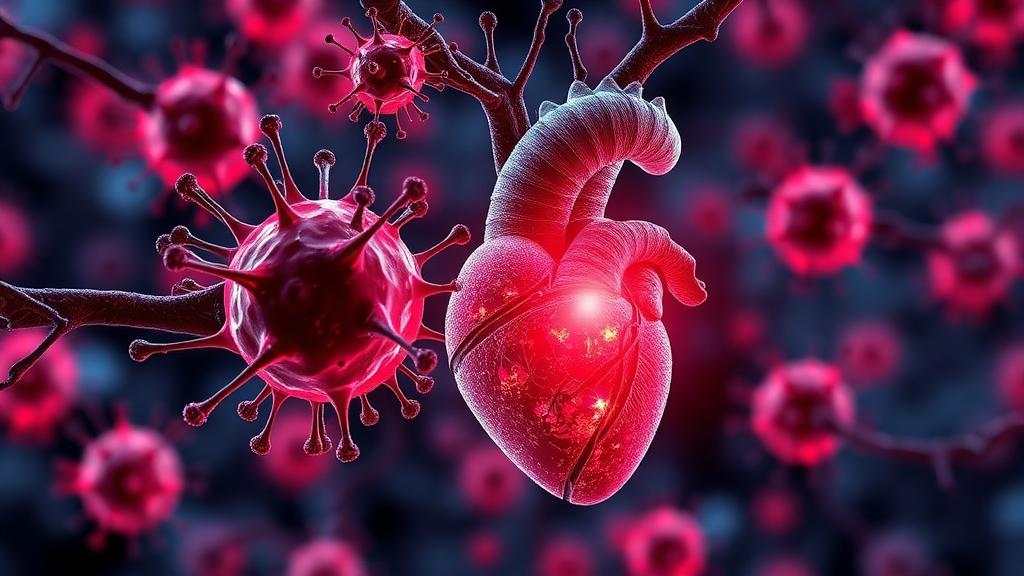
Image Credits: AI Generated
DOI: 10.1186/s12967-026-09052-7
Keywords: MAMs, myocardial ischemia-reperfusion injury, ferroptosis, lipid metabolism, mitochondria, endoplasmic reticulum, ventricular remodeling, PERK, reactive oxygen species, cardioprotection, GPX4, therapeutic targets
News Source: Ophelia Keating. (October 7, 2026). Cellular Contact Points Called MAMs Emerge as Central Players in Heart Attack Damage. Scienmag.