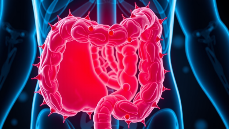
Colon cancer remains one of the deadliest malignancies worldwide, ranking as the second most common cause of cancer-related death and the third most prevalent cancer overall. Despite decades of progress in oncology, treatment still relies heavily on surgical resection and conventional chemotherapy, approaches that suffer from limited selectivity, harsh side effects, and effectiveness that is often confined to early-stage disease. A newly published review in Holistic Integrative Oncology argues that a radically different strategy, borrowed from biology itself, may finally change that trajectory. The review, led by Karan Kumar Dharme and colleagues at Rungta College of Pharmaceutical Sciences and Research in India, synthesizes the rapidly expanding field of biomimetic nanomaterials: engineered particles that cloak themselves in biological materials to slip past the body’s defenses and deliver drugs precisely where they are needed.
The core idea behind biomimetic nanomedicine is deceptively simple. Nanoparticles, whether synthetic or natural in origin, are coated with materials derived from living cells, such as membranes from platelets, macrophages, neutrophils, natural killer cells, erythrocytes, or even cancer cells themselves. Alternatively, they can be decorated with naturally derived biomolecules like monoclonal antibodies, viral capsids, and natural proteins, or with synthetic analogues such as targeting peptides and aptamers. This biological disguise confers several decisive advantages. The particles circulate in the bloodstream for longer periods, evade recognition by the reticuloendothelial system, accumulate preferentially in tumor tissue, and provoke far fewer unwanted immune reactions than conventional nanocarriers. In effect, the drug carrier stops looking like a foreign invader and starts looking like something the body already tolerates.
The review grounds this technological optimism in a detailed account of colon cancer biology. The disease typically unfolds over ten to twenty years through the well-characterized adenoma-carcinoma sequence. Loss of the APC tumor suppressor gene triggers uncontrolled proliferation and the formation of benign polyps. Subsequent activation of the KRAS oncogene drives growth into a large adenoma, while deletion of the DCC gene, involved in cell adhesion and apoptosis, permits abnormal cells to invade neighboring tissue. The final transformation comes with loss of p53, the guardian of DNA repair and programmed cell death, after which cells accumulate further mutations, breach the basement membrane, and metastasize to organs such as the liver and lungs. Understanding this stepwise molecular progression, the authors argue, is essential for designing nanocarriers that intervene at the right biological moment.
Among the most striking platforms described in the review are cell membrane-coated nanoparticles. By wrapping a synthetic drug-loaded core in the membrane of a red blood cell, platelet, or immune cell, researchers inherit the surface chemistry of that cell, including its complement-regulating proteins and adhesion molecules. Preclinical work highlighted in the review illustrates the potential vividly. In a mouse model of inflammation-driven colorectal cancer, biomimetic PLGA nanoparticles coated with red blood cell membranes and loaded with the enzyme shikimokinase produced stable body weights, reduced disease activity scores, longer colons, fewer tumor nodules, restored crypt architecture on histology, and enhanced apoptosis of tumor cells as measured by TUNEL staining. The membrane-coated formulations outperformed their uncoated counterparts across every endpoint, a result the authors present as evidence that biomimicry is not merely cosmetic but functionally therapeutic.
Exosome-based carriers represent a second major pillar. Exosomes are naturally occurring extracellular vesicles that cells use to communicate, and their innate ability to cross biological barriers makes them attractive delivery vehicles. Yet the review is candid about their limitations. Isolation methods such as ultracentrifugation, size-exclusion chromatography, and immunoaffinity capture yield heterogeneous populations with inconsistent purity, undermining reproducibility. Cargo loading through passive incubation, electroporation, or chemical transfection still suffers from low encapsulation rates and rapid release. Once injected, exosomes face rapid clearance, aggregation, and degradation in circulation. Engineering strategies, including genetic modification of donor cells to display tumor-targeting peptides on the exosome surface, offer improved intrinsic targeting, while surface functionalization provides greater flexibility and scalability. The authors call for systematic studies of drug loading, release kinetics, and therapeutic efficacy to establish the standardized protocols that clinical translation will demand.
Inorganic and metal-based nanoparticles form a third, chemically diverse family. Gold nanoparticles, ranging from one to one thousand nanometers and available in spherical, rod, star, and cage morphologies, can convert near-infrared light into heat, making them potent agents for photothermal therapy. In one study cited in the review, photostable gold nanoparticles averaging roughly 180 nanometers were absorbed by colon cancer cells, as confirmed by increased cellular granularity, and produced a dramatic drop in cell viability after treatment. Silver nanoparticles take a different route, generating reactive oxygen species that induce oxidative stress, mitochondrial damage, membrane rupture, and apoptosis. Research on the SW480 colon cancer cell line showed that silver nanoparticles functionalized with glutamine and conjugated with thiosemicarbazide triggered apoptosis through notable caspase activation and downregulation of the HULC long non-coding RNA and the PPFIA4 oncogene. Silver nanoparticles can also enhance the tumor accumulation of conventional drugs like doxorubicin while reducing toxicity to healthy tissue, and their strong light-scattering properties make them useful as contrast agents in dark-field microscopy and surface-enhanced Raman scattering.
The review also catalogs the contributions of softer organic platforms. Dendrimers, tree-like branched macromolecules whose size and surface chemistry are controlled generation by generation, can be loaded with drugs in their interior cavities or conjugated to their periphery. Fourth-generation PAMAM dendrimers carrying capecitabine shrank tumors in mouse models with fewer effects on blood and liver than the free drug, while pegylated PAMAM dendrimers decorated with AS1411 anti-nucleolin aptamers showed heightened sensitivity against HT29 and C26 colorectal cancer cells and efficacy in tumor-bearing mice. A fifth-generation L-lysine dendrimer modified with polyoxazoline improved the therapeutic index of SN-38, the active metabolite of irinotecan, by controlling drug release and extending circulation time. Liposomes, the oldest clinical nanomedicine platform, continue to evolve: FDA-approved formulations such as Doxil and DaunoXome established the field in the 1990s, and temperature-sensitive liposomal doxorubicin, known as Thermodox, delivered twenty-five times more drug to cancer cells than standard intravenous delivery in preclinical testing for colorectal liver metastases.
Carbon nanotubes and quantum dots round out the arsenal. Carbon nanotubes, cylindrical rolls of hexagonally bonded carbon atoms a few nanometers in diameter, penetrate plasma membranes readily thanks to their needle-like shape and vast surface area. Combining single-walled carbon nanotubes with the TRAIL protein increased the death rate in colorectal cancer cell lines roughly tenfold, and carboxylic acid functionalization exploits the acidic lysosomal environment of cancer cells, around pH 5.5 versus 7.4 in healthy tissue, to trigger pH-dependent drug release. Quantum dots, semiconductor particles smaller than ten nanometers, offer exceptional fluorescence and photochemical stability for bioimaging, and carbon quantum dots are emerging as diagnostic and therapeutic tools in their own right. The review notes that colon cancers frequently overexpress placenta-specific protein 1, providing a molecular anchor for targeted nanoparticle binding.
The translational picture is genuinely encouraging but far from settled. The review catalogs a growing roster of clinical trials involving nanotechnology in colon and gastrointestinal cancers, including studies of cetuximab-loaded nanoparticles, liposomal irinotecan combined with FOLFIRI and bevacizumab, liposomal mitomycin-C with capecitabine, and nanoliposomal irinotecan paired with TAS-102. Yet the authors are clear that major bottlenecks remain: scalable manufacturing, batch-to-batch reproducibility, and demanding regulatory pathways under FDA and EMA frameworks. Their proposed solutions include cost-effective microfluidic production, automated bioreactors, comprehensive protocols for exosome isolation and nanoparticle characterization, and early harmonization with regulatory safety and quality standards. Looking forward, they envision theranostic platforms that merge diagnosis and treatment in a single particle, biomimetic carriers that ferry gene-editing tools alongside chemotherapy, nanomaterials engineered to reprogram the tumor microenvironment, and the encapsulation of plant-derived phytochemicals to overcome their poor solubility and low bioavailability. If interdisciplinary collaboration can close the gap between laboratory proof-of-concept and clinical practice, the authors conclude, biomimetic nanomaterials could move colon cancer therapy decisively closer to being safer, more effective, and personally tailored.
Subject of Research: Biomimetic nanomaterials for targeted drug delivery in colon cancer
Article Title: Advances in biomimetic nanomaterials for targeted drug delivery in colon cancer
Article References: Dharme, K. K., Jain, P., Verma, G., & Uddin, A. (2026). Advances in biomimetic nanomaterials for targeted drug delivery in colon cancer. Holistic Integrative Oncology, 5(1), Article 58. https://doi.org/10.1007/s44178-026-00279-4
Image Credits: AI Generated
DOI: 10.1007/s44178-026-00279-4
Keywords: colon cancer, biomimetic nanoparticles, drug delivery, exosomes, cell membrane coating, gold nanoparticles, silver nanoparticles, dendrimers, liposomes, carbon nanotubes, quantum dots, nanomedicine
Cite Scienmag News
APA MLA Chicago
Nathaniel Bowman. (September 25, 2026). Cell-Disguised Nanoparticles Show Promise for Targeted Colon Cancer Therapy. Scienmag. https://scienmag.com/cell-disguised-nanoparticles-show-promise-for-targeted-colon-cancer-therapy/
Nathaniel Bowman. “Cell-Disguised Nanoparticles Show Promise for Targeted Colon Cancer Therapy.” Scienmag, 25 September 2026, https://scienmag.com/cell-disguised-nanoparticles-show-promise-for-targeted-colon-cancer-therapy/. Accessed 25 September 2026.
Nathaniel Bowman. “Cell-Disguised Nanoparticles Show Promise for Targeted Colon Cancer Therapy.” Scienmag. September 25, 2026. https://scienmag.com/cell-disguised-nanoparticles-show-promise-for-targeted-colon-cancer-therapy/
Copy citation Download RIS
Tags: biological material coatingbiomimetic nanomaterialsbiomimetic nanomedicinebiomimetic nanoparticlesbiomolecule functionalizationCancer drug deliverycarbon nanotubescell membrane coatingcell-disguised nanoparticlescolon cancerdendrimersDrug deliveryexosomesgold nanoparticlesimmune system evasion in nanomedicineinnovative cancer therapeutic strategiesLiposomesnanocarriers for cancer treatmentNanomedicinenanoparticle cloaking techniquesprecision oncologyquantum dotssilver nanoparticlestargeted colon cancer therapy