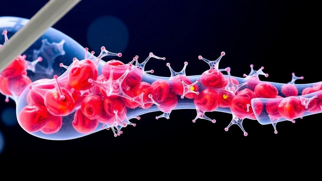
Every second, millions of cells in the human body die on purpose. Apoptosis, the highly choreographed program of cell suicide, has long been viewed as an ending: the cell shrinks, fragments, and is quietly cleared away by professional scavengers. But a growing body of research suggests that the dying cell does not simply vanish. As it disassembles, it releases a family of membrane-wrapped packages known as apoptotic extracellular vesicles, or ApoEVs, and these packages are turning out to be anything but inert debris. A new review published in the Journal of Translational Medicine by Shuwen Wu, Zhiqing Chen and colleagues at Guangzhou Medical University assembles the evidence that ApoEVs act as genuine signaling agents in the skeleton, orchestrating the constant demolition and reconstruction cycle known as bone remodeling.
Bone is not the static scaffold it appears to be on an X-ray. Throughout life, the skeleton undergoes continuous renewal through the coordinated activity of bone-forming osteoblasts, bone-resorbing osteoclasts, mechanosensing osteocytes, mesenchymal stem cells, immune cells and endothelial cells lining the vasculature. When this balance tips, the consequences are familiar and debilitating: osteoporosis, delayed fracture healing, and failed bone grafts. The Guangzhou team argues that ApoEVs sit at a previously underappreciated control point in this system, ferrying RNA molecules, proteins and lipids between the cell types that build and break down bone, and thereby shaping the communication networks that keep skeletal tissue in homeostasis.
Technically, ApoEVs are a heterogeneous category. The review distinguishes several subtypes based on size and biogenesis, including large apoptotic bodies, apoptotic microvesicles shed from the plasma membrane, apoptotic exosome-like vesicles, and smaller apoptotic small extracellular vesicles. What unites them is their origin in programmed cell death and their cargo: they encapsulate messenger RNAs, microRNAs, long noncoding RNAs and ribosomal RNAs, together with proteins and lipids that often reflect the identity of the parent cell. A vesicle released by a bone marrow mesenchymal stem cell carries a different molecular inventory than one shed by a mature osteoclast, and that identity matters for the message the vesicle delivers to its recipient.
Two signaling circuits dominate the review’s mechanistic analysis. The first is the RANKL/RANK/OPG axis, the central lever of osteoclast biology. RANKL, expressed on osteoblasts and osteocytes, binds RANK on osteoclast precursors and drives their differentiation into bone-resorbing giants, while osteoprotegerin acts as a decoy receptor that dampens the process. ApoEVs can modulate this axis from multiple directions, influencing the ratio of RANKL to OPG in the local microenvironment and thereby tuning how aggressively bone is broken down. The second circuit is the Wnt/β-catenin pathway, the master switch for osteoblast differentiation. When Wnt ligands engage their receptors, β-catenin accumulates and translocates to the nucleus, switching on genes such as RUNX2 and Osterix that commit stem cells to the bone-forming lineage. ApoEVs have been shown to carry Wnt pathway components and microRNAs that either amplify or suppress this cascade depending on their cargo.
The cargo story is where the biology becomes genuinely striking. In preclinical studies summarized by the review, apoptotic vesicles derived from mesenchymal stem cells delivered microRNAs that activated the AKT and ERK signaling cascades in recipient cells, promoting osteogenic differentiation marked by elevated alkaline phosphatase activity and expression of osteocalcin and bone sialoprotein. Vesicles from other sources carried insulin-like growth factor binding proteins, bone morphogenetic protein pathway components, or stanniocalcin 1, each steering recipient cells along distinct trajectories. Even the surface of the vesicle participates: the abundant phosphatidylserine on the outer leaflet of apoptotic membranes is recognized by receptors such as MerTK and TIM-4 on phagocytes, linking vesicle uptake to the same engulfment machinery that clears dying cells during normal tissue turnover.
Immune regulation emerges as a third pillar. Macrophage-derived apoptotic vesicles and T cell-derived vesicles can shift the inflammatory tone of the bone microenvironment, and the review highlights interactions with the STING pathway, a cytosolic DNA sensor that drives inflammatory signaling when activated. By modulating tumor necrosis factor alpha, interleukins and cyclooxygenase-2 signaling, ApoEVs appear to help resolve inflammation after injury, creating a permissive environment for regeneration. This osteoimmunological dimension matters clinically, because excessive inflammation is a hallmark of osteoporotic bone loss and a major obstacle to fracture healing and implant integration.
The translational implications are already being tested in animal models. In studies of critical-sized bone defects, scaffolds loaded with mesenchymal stem cell-derived ApoEVs accelerated regeneration, with the vesicles promoting both new bone formation and angiogenesis through vascular endothelial growth factor signaling. In ovariectomized rodents, the standard model of postmenopausal osteoporosis, ApoEV-based treatments improved bone microarchitecture and partially reversed the resorption-heavy imbalance. Vesicles derived from dental pulp stem cells and periodontal ligament stem cells have shown promise in periodontal and craniofacial repair, while platelet-derived and red blood cell-derived apoptotic vesicles have been explored for their regenerative and immunomodulatory properties. Mature osteoclast-derived vesicles, intriguingly, appear to feed back on osteoblasts, suggesting that even the bone-destroying cells contribute building materials to the renewal cycle.
What makes ApoEVs attractive as therapeutic vehicles is structural as much as chemical. Their lipid bilayer protects fragile RNA cargo from degradation by serum nucleases, their phosphatidylserine-rich surface promotes uptake by target cells, and their size allows them to penetrate the extracellular matrix of bone tissue. The review describes engineering strategies that exploit these properties: surface functionalization with the aspartic acid-serine-serine peptide to confer bone-targeting ability, incorporation into gelatin methacryloyl hydrogels or poly(lactic-co-glycolic acid) scaffolds for local, sustained delivery, and preconditioning of parent cells with agents such as strontium or 1,25-dihydroxyvitamin D3 to load vesicles with pro-osteogenic cargo before they are harvested.
Significant hurdles remain before ApoEVs reach the clinic. The review notes that standardization of isolation and characterization protocols is still immature, that the heterogeneity of vesicle subpopulations complicates dose definition, and that scalable production under good manufacturing practice conditions has yet to be demonstrated for skeletal applications. Questions about biodistribution, immunogenicity across donors, and the long-term fate of vesicle cargo in recipients also await rigorous answers. The authors frame these as the field’s next frontier rather than disqualifying obstacles, pointing out that the same challenges confronted and were largely resolved for the broader extracellular vesicle field now advancing through early clinical trials.
The conceptual shift, however, may prove as important as any therapy. For decades, apoptosis was taught as a one-way street ending in silent disposal. The Guangzhou review makes the case that the dying cell’s final act is communicative: it packages its regulatory contents into vesicles that instruct neighboring stem cells, immune cells and vascular cells on how to rebuild. In the skeleton, where the stakes include osteoporosis affecting hundreds of millions of people worldwide, that reframing turns cellular death from a passive endpoint into an active resource, and it positions apoptotic extracellular vesicles as candidates for a new generation of regenerative treatments built from the body’s own demolition byproducts.
Subject of Research: Roles of apoptotic extracellular vesicles in bone remodeling and regeneration
Article Title: Emerging roles of apoptotic extracellular vesicles in bone remodeling: mechanisms and applications
Article References: Wu, S., Chen, Z., Yuan, Q., Chen, H., Zhang, B., & Lin, T. (2026). Emerging roles of apoptotic extracellular vesicles in bone remodeling: mechanisms and applications. Journal of Translational Medicine. https://doi.org/10.1186/s12967-026-08921-5
Image Credits: AI Generated
DOI: 10.1186/s12967-026-08921-5
Keywords: apoptotic extracellular vesicles, bone remodeling, osteoporosis, bone regeneration, RANKL/RANK/OPG axis, Wnt/beta-catenin pathway, mesenchymal stem cells, osteoblasts, osteoclasts, osteoimmunology, extracellular vesicles, bone tissue engineering
News Source: Ophelia Keating. (October 5, 2026). Cell Death Debris Turns Out to Be a Master Regulator of Bone Renewal. Scienmag.