Cyclin-dependent kinase 4, long celebrated as the molecular accelerator that pushes cells through the cell cycle, has now been caught doing a second, very different job. In triple-negative breast cancer cells, CDK4 keeps the cellular skeleton from tearing itself apart, according to a study published in EMBO Reports. When the kinase is deleted or blocked, the cells swell, lose their polarity, sprout excessive finger-like filopodia, and eventually suffer a collapse of their vimentin intermediate filament network, with mitochondria crowding around the nucleus. The root cause, the researchers report, is a previously uncharacterized phosphorylation site on a protein called Myo9b, which CDK4 tags at serine 1935 to keep a RhoA-driven contractility program in check.
The team, led by Kanishka Parashar, Sarah Geller and senior author Lluis Fajas at the University of Lausanne, together with collaborators in Switzerland and elsewhere, set out to understand whether CDK4’s influence on the cytoskeleton was more than a side effect of its role in cell division. They worked with MDA-MB-231 cells, an aggressive triple-negative breast cancer line, in which the CDK4 gene had been knocked out using CRISPR-Cas9. Compared with wild-type controls, the knockout cells were significantly larger, with increased area and perimeter, and their surfaces were measurably rougher and taller when scanned with a technique called scanning ion conductance microscopy. The cells also sprouted membrane ruffles that projected upward from the surface, a sign of disorganized architecture. Reintroducing CDK4 reversed the size defect, and a pharmacological inhibitor of CDK4 and CDK6, palbociclib, produced a milder version of the same phenotype in wild-type cells.
Crucially, the knockout cells did not show a major shift in cell-cycle distribution, and their proliferation was only slightly reduced, which suggested that the cytoskeletal defects were not simply a consequence of slowed division. The researchers then turned to the actin cytoskeleton itself. In wild-type cells, F-actin was densely concentrated at the leading edge, giving the cells a clear front-rear polarity. In the knockout cells, actin was spread uniformly around the membrane, polarity was lost, and the cells retracted their edges more frequently, although the rate of edge expansion was not significantly different. The knockout cells also produced far more filopodia, thin actin-rich probes that cells use to sense their environment, and these were longer than normal. Restoring CDK4 expression, or silencing it in wild-type cells, produced the expected reciprocal effects, confirming that the kinase is required to keep the actin network polarized and balanced.
To find the molecular target, the investigators mined previously generated phosphoproteomic data from the same cell lines. Among the candidates whose phosphorylation dropped when CDK4 was absent was Myo9b, a Rho GTPase-activating protein that normally switches off RhoA, a master switch for actomyosin contractility. The serine at position 1935 sits within a canonical CDK4 recognition motif and had scored highly in kinase prediction analyses. In vitro, purified CDK4-cyclin D3 phosphorylated a recombinant fragment of Myo9b exclusively at that site, while a nearby serine at position 1926 remained untouched. The phosphorylation event, the authors conclude, is direct and specific.
What does that phosphorylation accomplish? Using the structure prediction tool AlphaFold3, the team modeled the Myo9b-RhoA complex with and without phosphorylation at the two candidate serines. Only when Myo9b carried a phosphate at S1935 did the RhoGAP domain of Myo9b come into close contact with RhoA, at inter-residue distances of roughly 4.5 to 6 angstroms. In the unphosphorylated state, and in models phosphorylated at S1926 alone or at both sites, the two proteins remained too far apart to interact. Proximity ligation assays in cells confirmed that the Myo9b-RhoA interaction was significantly reduced in CDK4 knockout cells, and immunoprecipitation of RhoA pulled down less Myo9b from the knockout lysates. The phosphorylation, in other words, appears to stabilize the very molecular handshake that allows Myo9b to restrain RhoA.
The downstream consequences were consistent with that model. CDK4 knockout cells showed significantly elevated RhoA activity and higher levels of phosphorylated myosin light chain, or pMLC, a readout of actomyosin contractility. Treating the knockout cells with the ROCK inhibitor Y-27632 brought pMLC back down to wild-type levels, confirming that the excess contractility was ROCK-dependent. Silencing CDK2 or CDK6 did not reproduce the effect, pointing to a specific role for CDK4 rather than a redundant function among related cyclin-dependent kinases. When the researchers reintroduced a phosphomimetic version of Myo9b, in which S1935 was replaced by glutamate to mimic a permanent phosphate, pMLC levels fell, front-rear polarity was partially restored, and filopodia numbers dropped. A phosphodead S1935A mutant, or a phosphomimetic at S1926, failed to rescue the phenotype, underscoring the specificity of the S1935 mark.
The cytoskeletal damage did not stop with actin. Vimentin, a load-bearing intermediate filament that helps the cell resist mechanical stress, reorganized dramatically in CDK4-deficient cells, collapsing into a compact perinuclear mass and losing its normal filamentous architecture. Phosphoproteomics also showed reduced vimentin phosphorylation and reduced total vimentin abundance in the knockout cells, although in vitro assays did not support direct phosphorylation of vimentin by CDK4, suggesting the effect is indirect. Because intermediate filaments help position organelles, the team examined mitochondria and found that they too collapsed into a perinuclear cluster in the knockout cells, mirroring the vimentin phenotype. The authors caution that they did not directly test whether the actomyosin changes cause the intermediate filament and mitochondrial defects, and that the two could be parallel consequences of CDK4 loss rather than a single linear pathway.
All of this architectural chaos had a behavioral cost. In scratch assays, CDK4 knockout cells closed the wound significantly faster than wild-type cells, and transwell migration assays confirmed that more knockout cells made it through the pores. But live-cell tracking revealed that the movement was erratic. Wild-type cells followed relatively straight, predictable paths, whereas knockout cells wandered, with broader deviation from a straight-line trajectory. Time-lapse imaging showed a distinctive contractility-driven migration cycle in the knockout cells, in which protrusions extended, formed contractile tails, and then snapped forward in rapid displacements. ROCK inhibition reduced the migration rate of the knockout cells, and reintroduction of the Myo9b-S1935E phosphomimetic mutant also slowed their migration, tying the behavioral change back to the phosphorylation site.
To see whether the mechanism operates beyond the dish, the researchers analyzed tumors grown from the same CDK4 wild-type and knockout cells implanted into the mammary fat pad of immunodeficient mice. Tumor sections from the knockout grafts showed significantly higher pMLC staining intensity and a greater proportion of pMLC-positive cells. Proteomic and transcriptomic analyses of those tumors revealed enrichment of cytoskeleton-associated pathways and Rho GTPase signaling, consistent with the in vitro findings. The authors note that the tumor microenvironment contains stromal and immune cells, so the pathway-level changes cannot be attributed exclusively to the cancer cells themselves.
Finally, the team looked for traces of the mechanism in human disease. In breast cancer tissue microarrays spanning stage I and stage II-III tumors, the proportion of CDK4-positive cells that also expressed pMLC, and the proportion of pMLC-positive cells that also expressed CDK4, both fell significantly in the more advanced tumors, suggesting a progressive uncoupling of CDK4 and contractility signaling during disease progression. In three large patient cohorts, SCAN-B, TCGA and the I-SPY2 trial dataset, CDK4 expression showed an inverse correlation with gene sets linked to RhoA signaling, actin organization and metastasis-associated processes, particularly in triple-negative and ER-negative tumors. The authors emphasize that these correlations are supportive rather than conclusive evidence of the mechanism. Taken together, the study positions CDK4 not merely as a cell-cycle engine but as a mechanical guardian of cellular architecture, one whose loss shifts triple-negative breast cancer cells toward a faster, less directed and more contractile state.
Subject of Research: A non-canonical role for CDK4 in maintaining cytoskeletal mechanical homeostasis in triple-negative breast cancer cells through phosphorylation of the RhoGAP Myo9b
Article Title: CDK4 maintains cytoskeletal homeostasis by restraining actomyosin contractility
Article References: Parashar, K., Geller, S., Ravera, A., Roger, C., Ambrosini, G., Drake, B. F., Shi, J., Prieto Baños, S., Stroh, S., Moi, D., Simo Riudalbas, L., Dessimoz, C., Fantner, G. E., Fasshauer, D., Ziegler, D. V., & Fajas, L. (2026). CDK4 maintains cytoskeletal homeostasis by restraining actomyosin contractility. EMBO Reports. https://doi.org/10.1038/s44319-026-00974-3
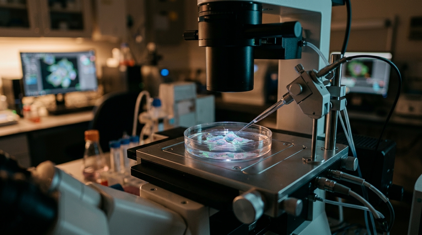
Image Credits: AI Generated
DOI: 10.1038/s44319-026-00974-3
Keywords: CDK4, Myo9b, RhoA, actomyosin contractility, triple-negative breast cancer, cytoskeleton, vimentin, phosphorylation, cell migration, palbociclib, pMLC, intermediate filaments
News Source: Nathaniel Bowman. (November 5, 2026). Cell-cycle kinase CDK4 keeps breast cancer cells mechanically stable, study finds. Scienmag.