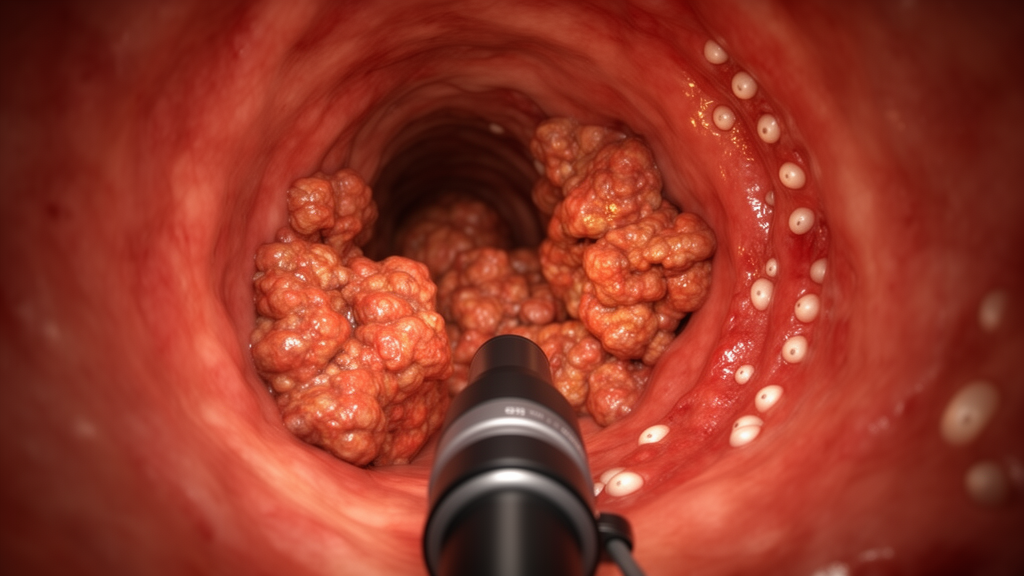
A 50-year-old Ethiopian man with rectal bleeding, lower abdominal pain, intermittent urination and weight loss was investigated for a rectal mass that appeared on imaging to be locally advanced cancer. A case report in BMC Infectious Diseases describes how repeat deep biopsies instead revealed pseudotumoral rectal schistosomiasis: eosinophil-rich granulomatous inflammation containing calcified Schistosoma mansoni ova, with no malignancy identified. The initial clinical assessment found a circumferential ulcerated rectal mass on colonoscopy, accompanied by narrowing of the rectal lumen.
Computed tomography and magnetic resonance imaging showed long-segment thickening of the rectal wall, invasion into the mesorectum, lymphadenopathy and encasement of both distal ureters. The imaging and endoscopic appearances therefore closely reproduced the pattern of a locally advanced rectal tumor. Laboratory findings supplied a different diagnostic clue.
The patient had leukocytosis, marked eosinophilia, microcytic anemia and renal impairment, while his carcinoembryonic antigen level was normal. Initial biopsy samples contained only necrotic and inflammatory tissue and did not establish a diagnosis. The report identifies the mismatch between the apparently malignant imaging appearance and the nondiagnostic superficial samples as an important feature of the case, with peripheral eosinophilia providing additional evidence for a parasitic inflammatory process.
Because the first specimens failed to reveal the cause of the mass, clinicians obtained repeat, deeper biopsies. Histological examination of the deeper samples showed eosinophil-rich granulomatous inflammation, calcified schistosome ova and no evidence of cancer. In the context of the clinical and radiological findings, these observations established pseudotumoral rectal schistosomiasis, a rare manifestation of chronic S. mansoni infection.
The condition is characterized by mass-forming granulomatous inflammation and fibrosis, allowing it to resemble colorectal carcinoma both clinically and radiologically. Identification of the ova in tissue provided the decisive evidence, demonstrating why adequate histopathological sampling was required to distinguish the inflammatory lesion from malignancy. The disease also had important consequences beyond diagnostic confusion.
Bilateral ureteric encasement progressed to obstructive uropathy and acute kidney injury, for which the patient required percutaneous nephrostomy and ureteric stenting. He was then treated with praziquantel and experienced partial recovery. As a single-patient case report, the study cannot establish the frequency of pseudotumoral rectal schistosomiasis, the usual response to treatment or the broader relationship between urinary obstruction and the disease.
Its scope is limited to the presentation, diagnostic course and immediate clinical outcome in one man. Nevertheless, the report shows that a parasitic inflammatory mass can closely mimic locally advanced rectal carcinoma while causing substantial renal and urinary complications, and that repeat deep biopsy can resolve the diagnostic uncertainty.
Subject of Research: A single-patient case report of pseudotumoral rectal schistosomiasis resembling advanced rectal carcinoma
Article Title: Pseudotumoral schistosomiasis mimicking locally advanced rectal carcinoma: a case report
Article References: Habte, Y. M., Habte, B. M., Areda, P. S., Fethudin, H. A., Hassen, K. A., Abdela, A. F., & Yimer, S. A. (2026). Pseudotumoral schistosomiasis mimicking locally advanced rectal carcinoma: a case report. BMC Infectious Diseases. https://doi.org/10.1186/s12879-026-14338-8
Image Credits: AI Generated
DOI: 10.1186/s12879-026-14338-8
Keywords: pseudotumoral rectal schistosomiasis, Schistosoma mansoni, rectal carcinoma mimic, case report, Eosinophilia, granulomatous inflammation, colorectal cancer diagnosis, obstructive uropathy, praziquantel, deep biopsy, Pseudotumoral, schistosomiasis
News Source: Ophelia Keating. (October 26, 2026). Case report identifies schistosomiasis mimicking advanced rectal cancer. Scienmag.