• HOME
  • NEWS
  • EXPLORE
    • CAREER
      • Companies
      • Jobs
    • EVENTS
    • iGEM
      • News
      • Team
    • PHOTOS
    • VIDEO
    • WIKI
  • BLOG
  • COMMUNITY
    • FACEBOOK
    • INSTAGRAM
    • TWITTER
Friday, January 16, 2026
BIOENGINEER.ORG
No Result
View All Result
  • Login
  • HOME
  • NEWS
  • EXPLORE
    • CAREER
      • Companies
      • Jobs
        • Lecturer
        • PhD Studentship
        • Postdoc
        • Research Assistant
    • EVENTS
    • iGEM
      • News
      • Team
    • PHOTOS
    • VIDEO
    • WIKI
  • BLOG
  • COMMUNITY
    • FACEBOOK
    • INSTAGRAM
    • TWITTER
No Result
View All Result
Bioengineer.org
No Result
View All Result
Home NEWS Science News Health

Carbon dots make calcium easier to track

Bioengineer by Bioengineer
November 12, 2019
in Health
Reading Time: 2 mins read
0
IMAGE
Share on FacebookShare on TwitterShare on LinkedinShare on RedditShare on Telegram

IMAGE

Credit: SIBET


In hospitals, doctors often advise patients to take calcium supplements. But does the calcium get into the cells that need it? Until recently, it’s been hard to tell.

But now, Prof. DONG Wenfei’s research group from the Suzhou Institute of Biomedical Engineering and Technology (SIBET) has developed a new type of fluorescent carbon dot that can effectively detect calcium levels in cells.

Calcium is essential to the life. Calcium ions regulate a variety of important cellular functions such as differentiation, proliferation, growth and gene transcription.

In addition, studies have reported that calcium status in cancer cells is associated with tumorigenesis, metastasis and angiogenesis. Other studies have also shown that abnormal changes in intracellular Ca2+ concentration are associated with the occurrence and development of Parkinson’s disease, Huntington’s disease and Alzheimer’s disease. Thus, detection of intracellular calcium levels is very important.

Conventional methods for calcium ion detection such as atomic absorption spectroscopy (AAS) and nuclear magnetic resonance (NMR) require expensive equipment and tedious sample preparation, limiting their applications in intracellular detection. Therefore, developing affordable, efficient, sensitive and simple techniques for detecting Ca2+ ions in cells is a priority. Fortunately, fluorescence detection meets these requirements.

As a new fluorescent probe, carbon dots (CDs) have been widely used in ion and small-molecule detection due to their extraordinary performance, which includes water solubility, excellent stability, nontoxicity, adjustable fluorescence and high quantum yield.

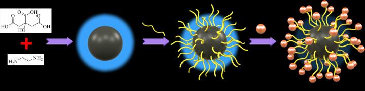
In this study, the researchers used an innovative approach to preparing functional CDs. They first synthesized the CDs from citric acid and ethylenediamine using a hydrothermal method. Then they modified the surface of the CDs with EGTA by reheating the solution.

This is a simple and convenient chemical preparation method, compared with the traditional cross-linking procedure, because all reactions are carried out in the same autoclave. The EGTA-modified CDs are the final product; no secondary purification and separation is required.

This work entitled “Two-Step Hydrothermal Preparation of Carbon Dots for Calcium Ion Detection” has been published on the ACS Applied Materials & Interface.

The CDs exhibit bright blue fluorescence. As calcium concentration increases, the fluorescence intensity drops sharply.

Cellular experiments show that CDs are nontoxic and have good biocompatibility, suggesting they may be a useful probe for intracellular Ca2+ detection.

In the future, researchers expect to develop fluorescent carbon dots in the near-infrared band with strong penetration. The distribution of calcium ions in internal organs could then be detected nondestructively.

###

Media Contact
XIAO Xintong
[email protected]

Original Source

http://english.cas.cn/newsroom/research_news/life/201911/t20191112_223119.shtml

Related Journal Article

http://dx.doi.org/10.1021/acsami.9b13737

Tags: cancerMedicine/HealthTechnology/Engineering/Computer Science
Share12Tweet8Share2ShareShareShare2

Related Posts

3D Printed Guides: Engineer-Surgeon Collaboration in Scoliosis

January 16, 2026

3D-Printed Implant Innovates Distal Femur Tumor Reconstruction

January 16, 2026

Assessing 3D Printing Accuracy in Total Sacrectomy

January 16, 2026

Revolutionary Support Program for Families of Cancer Patients

January 16, 2026
Please login to join discussion

POPULAR NEWS

  • Enhancing Spiritual Care Education in Nursing Programs

    155 shares
    Share 62 Tweet 39
  • PTSD, Depression, Anxiety in Childhood Cancer Survivors, Parents

    147 shares
    Share 59 Tweet 37
  • Robotic Ureteral Reconstruction: A Novel Approach

    77 shares
    Share 31 Tweet 19
  • Study Reveals Lipid Accumulation in ME/CFS Cells

    54 shares
    Share 22 Tweet 14

About

We bring you the latest biotechnology news from best research centers and universities around the world. Check our website.

Follow us

Recent News

3D Printed Guides: Engineer-Surgeon Collaboration in Scoliosis

Co-Infused Porous Carbon Enhances Polysulfide Management in Batteries

3D-Printed Implant Innovates Distal Femur Tumor Reconstruction

Subscribe to Blog via Email

Enter your email address to subscribe to this blog and receive notifications of new posts by email.

Join 71 other subscribers
  • Contact Us

Bioengineer.org © Copyright 2023 All Rights Reserved.

Welcome Back!

Login to your account below

Forgotten Password?

Retrieve your password

Please enter your username or email address to reset your password.

Log In
No Result
View All Result
  • Homepages
    • Home Page 1
    • Home Page 2
  • News
  • National
  • Business
  • Health
  • Lifestyle
  • Science

Bioengineer.org © Copyright 2023 All Rights Reserved.