• HOME
  • NEWS
  • EXPLORE
    • CAREER
      • Companies
      • Jobs
    • EVENTS
    • iGEM
      • News
      • Team
    • PHOTOS
    • VIDEO
    • WIKI
  • BLOG
  • COMMUNITY
    • FACEBOOK
    • INSTAGRAM
    • TWITTER
Tuesday, September 30, 2025
BIOENGINEER.ORG
No Result
View All Result
  • Login
  • HOME
  • NEWS
  • EXPLORE
    • CAREER
      • Companies
      • Jobs
        • Lecturer
        • PhD Studentship
        • Postdoc
        • Research Assistant
    • EVENTS
    • iGEM
      • News
      • Team
    • PHOTOS
    • VIDEO
    • WIKI
  • BLOG
  • COMMUNITY
    • FACEBOOK
    • INSTAGRAM
    • TWITTER
No Result
View All Result
Bioengineer.org
No Result
View All Result
Home NEWS Science News Health

Bright spot analysis for photodynamic diagnosis of brain tumors using confocal microscopy

Bioengineer by Bioengineer
April 11, 2019
in Health
Reading Time: 2 mins read
0
IMAGE
Share on FacebookShare on TwitterShare on LinkedinShare on RedditShare on Telegram

IMAGE

Credit: Kanazawa University

[Background]

Photodynamic diagnosis using 5-aminolevulinic acid (5-ALA) is now widely used for neurosurgical resection of brain tumors. Distinguishing a tumor from healthy tissue is based on greater 5-ALA-derived protoporphyrin IX accumulation in glioma cells than in non-cancerous cells, resulting in much greater red fluorescence (peak at 635 nm) when excited at 405 nm. However, it is still difficult to precisely distinguish the tumor margin and infiltrating regions from non-tumor tissue because the fluorescent boundary is usually vague. In our previous study, we noticed that bright spots in confocal microscopy images may be able to distinguish tumors from normal tissue.

[Methods]

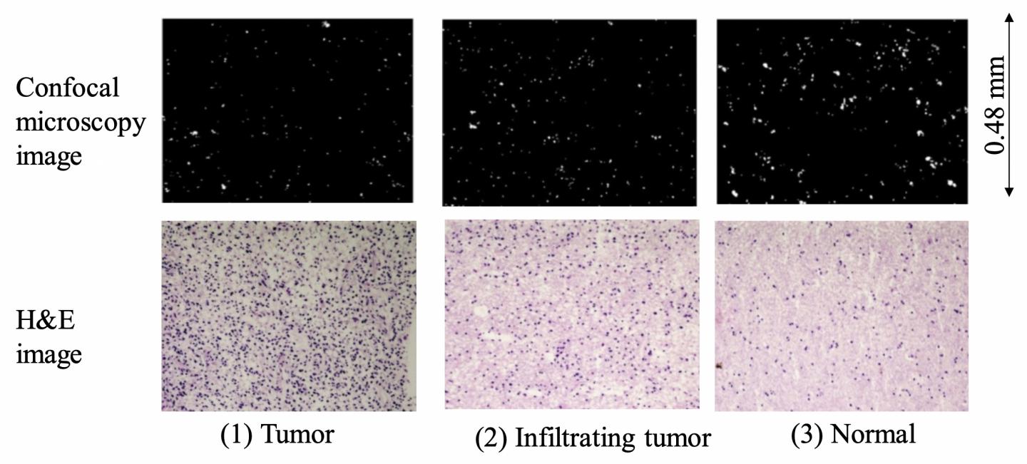
Brain tumor tissues resected from 5-ALA-treated patients was sectioned to evaluate bright spots captured by a 544.5-619.5 nm wavelength band-pass filter that eliminated the fluorescence induced by 5-ALA under a confocal microscope. Boarder regions and adjacent normal tissues were observed. Pathological inspection was performed to confirm the locations of tumors, infiltrating tumor cells, and normal tissue regions by hematoxylin and eosin (H&E) staining of serial sections of the same samples. Bright spot areas were measured in the same region used for pathological inspection. This method was applied to brain tumors with and without red fluorescence as well as glioblastoma (GBM) and non-GBM brain tumors.

[Results]

The bright spot area was substantially smaller in the GBM tumor than in normal brain tissues. It was also smaller in infiltrating tumors than in normal tissue at the margin. The same bright spot pattern was observed in tumors tissues without red fluorescence and in non-GBM tumors. Bright spot fluorescence has been suggested to be derived from lipofuscin based on emission spectra (mainly within 544.5?619.5 nm) and an optimal excitation wavelength (about 405 nm).

[Significance and future prospects]

Bright spot analysis is useful to facilitate discrimination of an infiltrating tumor from bordering normal tissue in photodynamic diagnosis using 5-ALA. This method is also potentially useful for tumors without 5-ALA-derived red fluorescence and non-GBM tumors. The mechanism of bright spot fluorescence reduction in tumors and its application for precise discrimination of brain tumors should be investigated further.

###

Media Contact
Tomoya Sato
[email protected]

Related Journal Article

http://dx.doi.org/10.1016/j.pdpdt.2019.02.005

Tags: Biomedical/Environmental/Chemical EngineeringcancerDiagnosticsMedicine/HealthSurgery
Share13Tweet8Share2ShareShareShare2

Related Posts

Vaccination Timing and Coverage Shape Measles Elimination

September 30, 2025

Enhancing Nursing Education with BOPPPS Model

September 30, 2025

Integrating Strongyloides Diagnostics into Rwanda’s Helminth Control

September 30, 2025

Evidence Use in Australian Clinical Networks Explained

September 30, 2025
Please login to join discussion

POPULAR NEWS

  • New Study Reveals the Science Behind Exercise and Weight Loss

    New Study Reveals the Science Behind Exercise and Weight Loss

    87 shares
    Share 35 Tweet 22
  • Physicists Develop Visible Time Crystal for the First Time

    73 shares
    Share 29 Tweet 18
  • How Donor Human Milk Storage Impacts Gut Health in Preemies

    59 shares
    Share 24 Tweet 15
  • Scientists Discover and Synthesize Active Compound in Magic Mushrooms Again

    56 shares
    Share 22 Tweet 14

About

We bring you the latest biotechnology news from best research centers and universities around the world. Check our website.

Follow us

Recent News

Vaccination Timing and Coverage Shape Measles Elimination

Future Neonatology: Boosting Interprofessional Collaboration Urged

Enhancing Nursing Education with BOPPPS Model

Subscribe to Blog via Email

Enter your email address to subscribe to this blog and receive notifications of new posts by email.

Join 60 other subscribers
  • Contact Us

Bioengineer.org © Copyright 2023 All Rights Reserved.

Welcome Back!

Login to your account below

Forgotten Password?

Retrieve your password

Please enter your username or email address to reset your password.

Log In
No Result
View All Result
  • Homepages
    • Home Page 1
    • Home Page 2
  • News
  • National
  • Business
  • Health
  • Lifestyle
  • Science

Bioengineer.org © Copyright 2023 All Rights Reserved.