A groundbreaking advancement in brain imaging heralds a new era in the early detection of Alzheimer’s disease, uncovering pathological tau proteins well before clinical symptoms arise. Researchers from the University of Pittsburgh School of Medicine have executed a comprehensive, multicenter study that compares two positron emission tomography (PET) tracers—Flortaucipir and MK6240—to determine their efficacy in identifying tau pathology, a pivotal biomarker closely linked to Alzheimer’s disease progression. The findings, published in the prestigious journal The Lancet, highlight the crucial implications for diagnosis, clinical trial recruitment, and future therapeutic strategies.
Alzheimer’s disease, a devastating neurodegenerative disorder, is characterized by the accumulation of amyloid plaques followed by tau protein tangles within the brain. While amyloid presence signals the initial stage of pathology, it is the aggregation of tau proteins that more directly correlates with neuronal dysfunction and cognitive decline. Despite this known hierarchy in pathological events, current clinical practices primarily rely on the FDA-approved tracer Flortaucipir, which detects advanced tau pathology but may lack sensitivity in early-stage disease detection.
The study, led by Dr. Tharick Pascoal, juxtaposed Flortaucipir with MK6240, a novel tracer currently used predominantly in research contexts. By administering paired tau PET scans to 682 cognitively diverse participants across multiple centers, the investigators ensured each individual was evaluated at the same disease timepoint using both tracers. This methodological rigor mitigated temporal variability and allowed a direct head-to-head assessment of tracer performance.
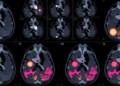
Results demonstrated that MK6240 significantly outperformed Flortaucipir in detecting tau pathology, particularly among cognitively unimpaired individuals who were amyloid-β positive. MK6240 identified tau positivity in 15% of this population, more than doubling the 6% detection rate observed with Flortaucipir. This enhanced sensitivity translates into detecting 23 additional tau-positive cases per 100 people scanned, potentially enabling earlier and more precise interventions to halt or slow disease progression on the cusp of symptom manifestation.
In participants already exhibiting cognitive impairment, MK6240 continued to surpass Flortaucipir, identifying tau involvement in 28% compared to Flortaucipir’s 16%. This difference corresponds to the identification of 15 additional cases of mild cognitive impairment and 21 more dementia cases per 100 individuals scanned. Such improvement in detection accuracy captures a more detailed snapshot of the neurodegenerative cascade, enabling clinicians to stage disease severity with far greater nuance.
Tau pathology’s central role in Alzheimer’s disease biology lies in its ability to instigate and propagate neurodegeneration. Studies have underscored that individuals harboring amyloid plaques without concomitant tau tangles seldom develop behavioral symptoms. Conversely, tau aggregation sets the stage for irreversible synaptic dysfunction and neuronal loss. Consequently, the early and accurate quantification of tau burden is indispensable not only for identifying at-risk individuals but also for refining clinical trial enrollment criteria and avoiding unnecessary treatment in patients unlikely to benefit.
The implications for this development extend beyond diagnostics into therapeutic decision-making. With the advent of disease-modifying therapies targeting amyloid and potentially tau, distinguishing which patients are truly on an Alzheimer’s trajectory becomes paramount. Employing a more sensitive tracer like MK6240 could optimize patient selection, enhance monitoring of treatment efficacy, and minimize exposure to costly interventions for those without significant tau pathology.
Researchers emphasize the importance of comprehensive cognitive assessments in tandem with imaging to contextualize biomarker findings within clinical symptomatology. The study design incorporated rigorous neuropsychological evaluations within a tight 45-day window of PET imaging, ensuring that imaging findings were directly correlated with cognitive status. This holistic approach underscores the value of integrating molecular imaging with clinical phenotyping for precision medicine.
While MK6240 shows remarkable promise, it is noteworthy that it remains unapproved by the FDA for routine clinical use, unlike Flortaucipir, which holds approval specifically for advanced tau detection. The current study’s robust evidence base lays the groundwork for potential regulatory approval and widens the horizon for clinical adoption. The improved detection capabilities suggest that MK6240 could soon transition from a research tool to a frontline diagnostic modality.
This research is part of a broader initiative funded by a $40 million award from the National Institutes of Health’s National Institute on Aging, aiming to standardize and harmonize tau PET imaging methodologies across centers globally. Such efforts are critical to ensure reproducibility, facilitate large-scale studies, and implement biomarker-driven approaches in routine clinical care.
The potential for MK6240 to redefine Alzheimer’s disease diagnostics exemplifies how cutting-edge tracers can reveal pathology earlier, ultimately shaping the future of therapeutic intervention timelines. As neuroimaging technology evolves, coupling molecular specificity with clinical utility remains the ultimate goal in combating this complex and multifaceted disease.
As the scientific community digests these pivotal findings, the clinical landscape may soon pivot toward earlier, more accurate Alzheimer’s diagnoses, with tau PET imaging playing a front-line role. Enhanced detection not only informs prognosis but may also catalyze a paradigm shift in how neurodegenerative diseases are approached—from reactive management to proactive prevention.
Continued collaborative efforts among neurologists, psychiatrists, radiologists, and molecular scientists will be vital to translate imaging innovations into real-world impact. This landmark study from the University of Pittsburgh reinforces that the future of Alzheimer’s diagnosis is not just in seeing, but in detecting earlier and staging more precisely—a game changer in the ongoing battle against this devastating disease.
Subject of Research: Alzheimer’s disease diagnosis using tau PET imaging tracers
Article Title: Comparison of [18F]flortaucipir and [18F]MK6240 for the detection of tau pathology in Alzheimer’s disease (HEAD): a multicentre, prospective, cross-sectional, within participant study
News Publication Date: 28-May-2026
Web References:
University of Pittsburgh School of Medicine: https://www.medschool.pitt.edu/
The Lancet Article: https://www.thelancet.com/journals/lancet/article/PIIS0140-6736(26)00417-4/fulltext
NIH Project Summary: https://reporter.nih.gov/search/u_IngbfG7kWbhhVnDzEFsw/project-details/10900738
HEAD Study: https://head-study.info/home
Pascoal Lab: https://pascoallab.org/
Dementia and Cognitive Disorders at UPMC: https://www.upmc.com/services/neurology/services/dementia-cognitive-disorders
Image Credits: UPMC
Keywords: Alzheimer’s disease, tau pathology, neuroimaging, PET scan, Flortaucipir, MK6240, tau PET tracers, amyloid-β, neurodegenerative diseases, early diagnosis, molecular imaging, clinical trials
Tags: Alzheimer’s clinical trial recruitmentAlzheimer’s disease brain imagingearly detection of Alzheimer’s tau proteinearly-stage tau aggregation detectionFlortaucipir vs MK6240 efficacymulticenter tau PET studyneurodegenerative disease diagnostic advancementsnovel PET tracers for Alzheimer’s diagnosistau pathology biomarkers in Alzheimer’stau protein and cognitive declinetau protein PET tracers comparisonUniversity of Pittsburgh Alzheimer’s research