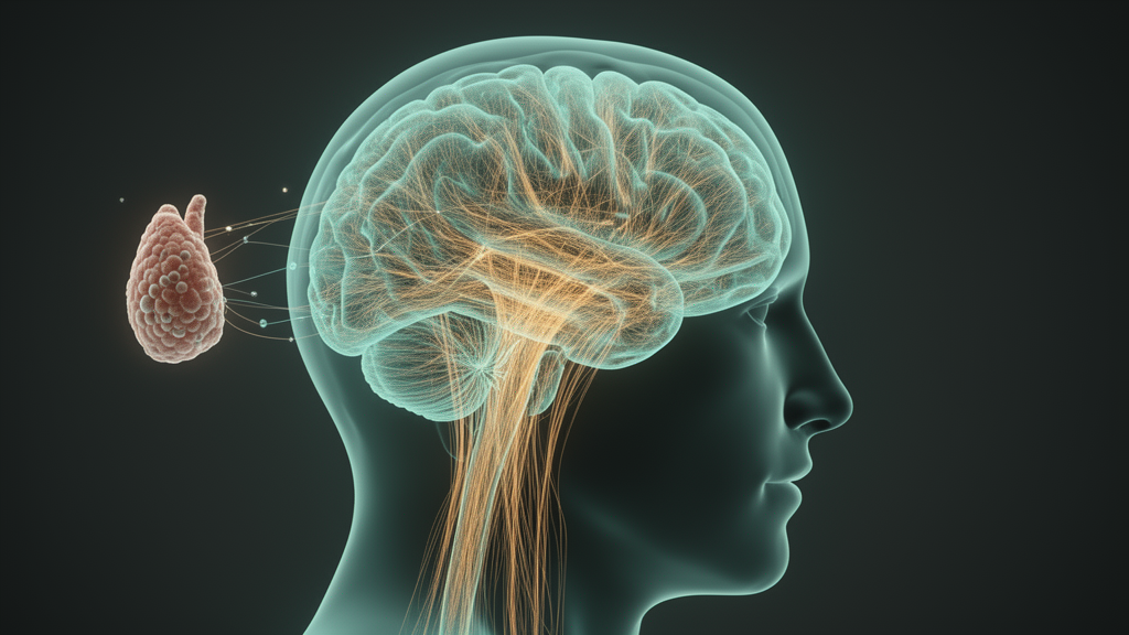
Depression is one of the most common and most underdiagnosed companions of cancer. In lung cancer in particular, depressive symptoms erode quality of life, complicate treatment decisions, and have been linked in meta-analyses to worse survival. Yet the biological signature of this comorbidity has remained almost entirely unmapped. A new exploratory neuroimaging study from Beijing now offers a first look at what happens to the brain’s intrinsic wiring when lung cancer patients also carry depressive symptoms, and the results point to a distributed pattern of altered functional connectivity that tracks with how severe those symptoms are.
The study, published in Supportive Care in Cancer, was led by Huixia Zhou, Wei Tang, Hengqin Ren and colleagues from the Institute of Psychology of the Chinese Academy of Sciences and the National Cancer Center/Cancer Hospital of the Chinese Academy of Medical Sciences. The team recruited 75 lung cancer patients and divided them into two groups: 33 patients with comorbid depressive symptoms and 42 age- and sex-matched patients without them. Depressive symptoms were assessed with the Patient Health Questionnaire-9, or PHQ-9, a nine-item screening instrument whose diagnostic accuracy as a depression screen has been validated in meta-analyses and whose Chinese version has established reliability in the general population.
The technical heart of the study is resting-state functional connectivity, or RS-FC. When a person lies still in an MRI scanner without performing any task, spontaneous low-frequency fluctuations of blood-oxygen-level-dependent signal ripple through the brain in correlated patterns. Regions whose fluctuations rise and fall together are considered functionally connected, and the resulting map reflects the brain’s intrinsic organization. The researchers processed their imaging data with DPARSF, a MATLAB toolbox for pipeline analysis of resting-state fMRI, and calculated whole-brain RS-FC for every participant. They then compared connectivity between the two patient groups to identify region pairs whose coupling differed with comorbid depressive symptoms.
The comparison revealed altered RS-FC across eight brain region pairs. These included the left calcarine cortex with right cerebellar lobules IV–V, the left medial frontal gyrus with the left anterior cingulate gyrus, the left insula with the left middle temporal gyrus, the left precentral gyrus with left cerebellar lobule III, the left thalamus with cerebellar vermis I and II, the right olfactory cortex with the right fusiform gyrus, the right rectus gyrus with right cerebellar lobule VIIb, and a further pair linking right cerebellar lobule VIIb with the right olfactory cortex. The pattern spans visual, frontal, insular, thalamic, olfactory and cerebellar territory, and notably involves the cerebellum repeatedly, a structure long associated with motor control but increasingly recognized as a hub for cognition and emotion.
Several of these region pairs make neurobiological sense in the context of depression. The anterior cingulate cortex shows well-documented functional connectivity changes in depression, and the anterior insula has been described as a gatekeeper of executive control, a system implicated in the cognitive regulation of negative emotion. Olfactory cortex involvement is also consistent with a growing literature linking impaired olfaction to depression, with olfactory function proposed as a potential cognitive marker of psychiatric disorders. The fusiform gyrus, meanwhile, has been reported to show hypoconnectivity with sensorimotor areas in major depressive disorder.
Beyond group differences, the team examined how connectivity related to symptom severity within the depressed group. Total PHQ-9 scores correlated negatively with RS-FC between multiple brain region pairs, and the correlations were strikingly strong. The strongest was between the left superior frontal gyrus and the right middle temporal gyrus, with a correlation coefficient of r = −0.72 (p < 0.001). The left superior frontal gyrus also correlated negatively with the left temporal pole middle temporal gyrus (r = −0.69), the left triangular inferior frontal gyrus correlated negatively with the same temporal pole region (r = −0.65), and the left medial superior frontal gyrus correlated negatively with the right middle temporal gyrus (r = −0.65). Two further pairs involved the cerebellar crus II: the left insula correlated negatively with right cerebellar crus II (r = −0.6), and the right superior temporal gyrus correlated negatively with the same cerebellar region (r = −0.67).
In plain terms, the more severe a patient’s depressive symptoms, the weaker the coupling between frontal regions that support cognitive control and temporal regions involved in language, social-signal processing and episodic memory retrieval. This fronto-temporal decoupling echoes findings from large-scale meta-analyses of resting-state network dysfunction in major depressive disorder, and the cerebellar crus II correlations align with earlier reports that altered cerebellar-cerebral connectivity can reliably identify major depressive disorder. The authors suggest these patterns may reflect disrupted top-down regulation of negative affect, though they are careful not to overclaim.
That caution is warranted, and the study is explicit about its limits. This was an exploratory, cross-sectional investigation of a heterogeneously treated Chinese lung cancer population. Data on psychotropic medication use, cancer-related symptoms such as pain and fatigue, and detailed treatment histories were not systematically collected. Because chemotherapy, persistent cancer-related fatigue, and cancer pain have each been shown in prior neuroimaging work to alter brain functional connectivity independently of mood, the observed patterns cannot be attributed specifically to depressive symptoms. The authors frame their findings as preliminary and hypothesis-generating rather than definitive markers of comorbid depression, and they note that no datasets were generated or analyzed during the current study in the sense of shared repositories.
The work was supported by grants from the Chinese National Programs for Brain Science and the National Natural Science Foundation of China, and it was approved by the Institutional Review Board of the Institute of Psychology, Chinese Academy of Sciences, with all participants providing written informed consent. The corresponding authors are Hongmei Zhang, Fengwei Tan and Xiangyang Zhang. The team calls for future longitudinal studies with systematic assessment of medication, treatment and multidimensional symptom burden to determine whether these connectivity patterns specifically reflect depressive symptoms or broader cancer-related sequelae. If they do, the findings could eventually support imaging-based identification of lung cancer patients at risk of depression, opening a window for earlier psychological intervention in a population where it is desperately needed.
Subject of Research: Neuroscience
Article Title: Altered resting-state functional connectivity in lung cancer patients with comorbid depressive symptoms
Article References: Zhou, H., Tang, W., Ren, H., Li, K., Ren, N., Li, R., Duan, J., Wang, D., Zhang, H., Tan, F., & Zhang, X. (2026). Altered resting-state functional connectivity in lung cancer patients with comorbid depressive symptoms. Supportive Care in Cancer, 34(11), Article 1114. https://doi.org/10.1007/s00520-026-11315-4
Image Credits: AI Generated
DOI: 10.1007/s00520-026-11315-4
Keywords: lung cancer, depression, resting-state fMRI, functional connectivity, PHQ-9, cerebellum, Supportive Care in Cancer, Altered, resting-state, functional, connectivity, lung
News Source: Nathaniel Bowman. (October 17, 2026). Brain Wiring Shifts Seen in Lung Cancer Patients With Depressive Symptoms. Scienmag.