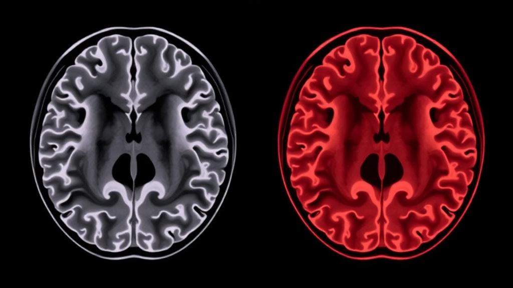
For millions of people with gluten sensitivity, the consequences of eating wheat, barley or rye extend far beyond the gut. Neurologists have long known that the immune reaction triggered by gluten can reach the brain, producing conditions with names that most of the public has never heard of: gluten ataxia, in which patients progressively lose their ability to walk steadily, and gluten encephalopathy, in which they struggle with a foggy, slowed mind and debilitating headaches. Now a team at Sheffield Teaching Hospitals and the University of Sheffield has used a sophisticated brain-imaging technique to show that these two disorders, although both driven by the same dietary trigger, leave distinctly different chemical fingerprints on the same structure — the cerebellum. The findings, published open access in the Journal of Neurology, offer some of the clearest evidence yet that the cerebellum is not merely a movement-control machine but a region deeply involved in thinking and emotion.
The technique at the heart of the study is proton magnetic resonance spectroscopy, often abbreviated as MR spectroscopy or MRS. Unlike conventional MRI, which produces detailed pictures of brain anatomy, MRS measures the concentrations of specific chemicals within a chosen volume of tissue. The researchers focused on one particular measurement: the ratio of N-acetyl-aspartate, usually shortened to NAA, to creatine, abbreviated Cr. NAA is a metabolite found almost exclusively inside healthy, functioning neurons, so a fall in NAA is widely interpreted as a sign of neuronal dysfunction or injury. Creatine, by contrast, is a stable metabolite whose levels remain essentially constant even in disease states. Dividing one by the other therefore yields a numerical index of neural health that is largely independent of the overall size or composition of the sampled region, making it an ideal yardstick for comparing patients with healthy volunteers.
The Sheffield group has spent years refining this approach in the context of gluten-related neurological disease. Patients referred to their dedicated gluten sensitivity and neurology clinic routinely undergo MRS of two cerebellar targets at baseline, before starting a gluten-free diet: the vermis, the narrow worm-shaped midline structure of the cerebellum, and the right cerebellar hemisphere. For the new analysis, the team retrospectively assembled spectroscopy data from 40 consecutive patients diagnosed with gluten ataxia and 40 consecutive patients diagnosed with gluten encephalopathy, deliberately excluding anyone who showed a mixture of the two conditions or who had evidence of gluten neuropathy. These were compared with normative data previously collected from 55 healthy adults. Diagnosis in both patient groups rested on the presence of circulating gluten-related antibodies — tissue transglutaminase 2, endomysial antibodies, antigliadin antibodies of the IgG or IgA class, or the more brain-specific transglutaminase 6 antibodies — together with the absence of any alternative explanation for the neurological symptoms.
The demographic picture that emerged was itself revealing. Patients with gluten ataxia had developed their neurological symptoms at a mean age of 50 years, whereas those with gluten encephalopathy had fallen ill at a mean age of just 37. Both groups had lived with their symptoms for an average of 11 years by the time of scanning, and the healthy controls had a mean age of 41 at the time of their scans. Because the encephalopathy group was actually younger than the control group, the researchers argue that age differences cannot plausibly explain the spectroscopic abnormalities they observed — a point that matters, since NAA levels can decline with normal ageing.
The headline result concerns where in the cerebellum each disease strikes. Patients with gluten ataxia showed a highly significant reduction in the vermis NAA/Cr ratio: 0.85 compared with 0.99 in healthy controls, a difference that reached a p-value of 0.0000105 using Welch’s t-test. In the same patients, the right hemisphere ratio of 0.969 versus 1.01 in controls was not statistically significant, with a p-value of 0.058. The pattern in gluten encephalopathy was almost the mirror image. These patients showed a significant reduction in the right hemisphere NAA/Cr ratio — 0.926 versus 1.01 in controls, p = 0.00053 — but no difference from controls in the vermis. A direct statistical comparison between the two patient groups confirmed the dissociation: vermis values differed dramatically between gluten ataxia and gluten encephalopathy, with p below 0.0001, while right hemisphere values did not differ significantly between the two groups.
What makes this dissociation scientifically satisfying is how neatly it maps onto the clinical pictures. The vermis is the part of the cerebellum most critically involved in coordinating gait and balance, and gait instability is the defining complaint of patients with gluten ataxia. Vermian involvement is also characteristic of other immune-mediated ataxias, so the spectroscopy findings place gluten ataxia squarely within that broader family of disorders. Patients with gluten encephalopathy, by contrast, typically do not complain of balance problems and show little evidence of ataxia on examination — yet their right cerebellar hemispheres are chemically abnormal. The researchers note that they could not assess the left hemisphere because their clinical protocol does not routinely sample it, but they point to existing evidence that lesions of the right cerebellar hemisphere produce greater cognitive deficits than lesions of the left.
That link between the right cerebellar hemisphere and cognition brings the study into one of the most exciting conceptual shifts in modern neuroscience: the recognition of the cerebellum as a cognitive organ. This rethinking owes much to the pioneering work of Schmahmann and colleagues, who described the cerebellar cognitive affective syndrome, sometimes called Schmahmann syndrome — a constellation of deficits in executive function, spatial cognition, language and emotional regulation that follows cerebellar injury. Assessment of the syndrome draws on some tasks familiar from general cognitive batteries such as the Montreal Cognitive Assessment, including phonemic fluency and delayed recall, but adds distinctive tests of category switching, inhibition and affect. The Sheffield team is careful to stress that gluten encephalopathy is not the same thing as cerebellar cognitive affective syndrome, but their data suggest that, in the context of gluten sensitivity, right hemisphere involvement may manifest primarily, and perhaps even exclusively, as cognitive limitation rather than as ataxia.
The clinical stakes are considerable because the symptoms of gluten encephalopathy can be devastating in everyday terms. Patients characteristically describe a foggy brain, slow mentation and an inability to think straight, often accompanied by reduced processing speed and intermittent migraine-like headaches, sometimes with unexplained sensory symptoms and, in a minority, white matter changes on conventional MRI that are often labelled vascular. These complaints can be severe enough to interfere with the ability to work. Yet the response to treatment can be striking: patients frequently report dramatic improvement on a strict gluten-free diet, with headaches often disappearing within weeks and cognitive clarity and sensory symptoms resolving over six months to a year. Crucially, the spectroscopy measure itself appears to track this recovery — the Sheffield group has previously demonstrated that the NAA/Cr ratio in patients with gluten ataxia improves after the introduction of a gluten-free diet, which means MRS can serve not only as a diagnostic tool but as an objective monitor of treatment response.
The new study also connects to a sobering recent finding from the same group about how common cerebellar cognitive dysfunction is in this population. Using a dedicated cognitive scale, the team found definite cerebellar cognitive affective syndrome in 39.2 percent of patients with neurological manifestations of gluten sensitivity attending their clinic, probable syndrome in a further 26.4 percent and possible syndrome in 18.4 percent, leaving only 16 percent free of such cognitive issues. Because most of those patients were already on a gluten-free diet — which improves both spectroscopy findings and clinical state — the true scale of the problem in untreated patients is likely to be substantially larger. The same study showed that persisting IgA transglutaminase 6 antibodies, a more sensitive marker of gluten-related neurological dysfunction, were associated with greater regional brain atrophy, along with more depression, greater symptom severity and poorer physical functioning.
Many questions remain. The researchers openly acknowledge that they cannot yet explain why some patients with gluten sensitivity present exclusively with ataxia while others present exclusively with encephalopathy, even though the cerebellum appears to be almost universally involved in gluten-related neurological disease. The pathophysiology of gluten encephalopathy, in particular, remains unresolved, especially given that only a small proportion of affected patients show white matter abnormalities on conventional MRI. What the spectroscopy data do establish is a principled framework: a single dietary trigger, a single vulnerable brain region, and two reproducible patterns of regional dysfunction that correspond to two distinct clinical syndromes. For patients, the practical message is potentially life-changing — a treatable, diet-responsive cause of both progressive unsteadiness and foggy thinking can be detected with a chemical scan of the cerebellum, and the earlier the gluten-free diet begins, the better the prospects for recovery.
Subject of Research: MR spectroscopy of cerebellar dysfunction in gluten ataxia and gluten encephalopathy
Article Title: MR spectroscopy of the cerebellum in gluten ataxia and gluten encephalopathy
Article References: Hadjivassiliou, M., Grunewald, R., Croal, I., & Hoggard, N. (2026). MR spectroscopy of the cerebellum in gluten ataxia and gluten encephalopathy. Journal of Neurology, 273(11), Article 658. https://doi.org/10.1007/s00415-026-14200-6
Image Credits: AI Generated
DOI: 10.1007/s00415-026-14200-6
Keywords: gluten sensitivity, gluten ataxia, gluten encephalopathy, cerebellum, MR spectroscopy, N-acetyl-aspartate, cerebellar vermis, cerebellar cognitive affective syndrome, gluten-free diet, transglutaminase 6 antibodies, neurology, coeliac disease
News Source: Cassandra Pierce. (October 13, 2026). Brain Scan Study Reveals How Gluten Can Attack the Cerebellum in Two Very Different Ways. Scienmag.