


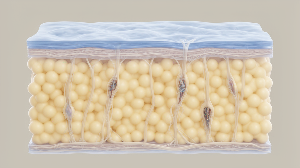
Lymphedema, the chronic swelling that follows damage to the lymphatic system, has long resisted a full explanation. Why do women develop it far more often than men? Why does primary lymphedema so often appear at puberty? Why has no laboratory reliably reproduced the disease in animals? A new hypothesis paper published in Frontiers in Physiology proposes a single anatomical answer that cuts across all of these puzzles: the amount of subcutaneous adipose tissue a person carries before lymphedema develops may determine whether, and how visibly, the condition manifests.
The paper, authored by Hiroo Suami of the Australian Lymphoedema Education, Research and Treatment Centre at Macquarie University and Atsushi Fujita of the University of Tokyo, is explicitly a hypothesis and theory article rather than a definitive clinical trial. The authors draw on imaging data from animals and humans, including indocyanine green lymphography performed at their clinic, to argue that baseline subcutaneous fat volume correlates with the amount of interstitial fluid the body produces, and therefore with the workload placed on the lymphatic system. If correct, subcutaneous adipose tissue could serve as a measurable parameter in clinical settings, something the field currently lacks.
Lymphedema is traditionally described as an imbalance between interstitial fluid production and lymphatic drainage capacity. When interstitial fluid production exceeds what the lymphatic system can clear, fluid accumulates in tissues. Yet, as the authors point out, no established method exists to quantify either of those volumes directly. Lymphedema is a subtype of chronic edema attributed to impaired lymphatic transport caused by structural or functional disorders of the lymphatic system, and it most commonly affects the extremities, though it can also arise in the head and neck after cervical lymph node dissection or in the breast after breast-conserving surgery. Clinically it is divided into secondary lymphedema, associated with surgery, trauma or other events causing lymphatic insufficiency, and primary lymphedema, which lacks any antecedent event, a classification based solely on medical history.
The starting point for the hypothesis is a comparative anatomy observation. Humans and pigs are so-called fixed-skin animals, in which the dermis is firmly connected to underlying tissues, while rodents, dogs and cats are loose-skin animals with movable skin and very thin layers of subcutaneous adipose tissue. The authors propose that thin subcutaneous fat in these species means lower interstitial fluid production, and therefore a higher threshold for lymphedema development. This, they argue, may explain why decades of attempts have failed to produce a reliable animal model reproducing persistent, human-like limb swelling accompanied by chronic adipose and fibrotic tissue remodeling.
The animal evidence is nuanced. In previous canine studies modeled on oncologic procedures such as lymph node dissection and radiation therapy, acute edema did occur after lymphatic injury, and lymphangiography demonstrated significant lymphatic structural changes resembling human lymphedema, including dermal backflow and lymphatic regeneration. However, the canine models failed to demonstrate persistent limb swelling associated with adipose hypertrophy. Rodent hind-limb models showed only transient swelling lasting approximately one month, and the widely used rodent tail model, while useful for physiological and pharmacological studies, has doubtful anatomical relevance to human limb lymphedema. The authors suggest that if researchers focused on lymphatic morphological changes rather than limb volume alone, these canine models might be considered successful representations of the human disease. They further propose that species with greater limb adiposity, such as swine or ovine species, may serve as more anatomically relevant models.
Clinical observations from the authors’ own clinic support the fat-volume idea from the human side. Obesity is a well-recognized risk factor for lymphedema, and it impairs multiple aspects of lymphatic function: it increases lymphatic vessel permeability, reduces the pumping rate of collecting vessels, and is associated with chronic inflammation and fibrosis. But lymphedema also occurs in lean individuals, and here the authors report a striking discrepancy. Using indocyanine green lymphography alongside standard circumferential tape or volume measurement, they observed lean patients whose imaging showed advanced lymphedema, at MD Anderson Cancer Center Stage 3 and 4, yet who had minimal limb enlargement. In overweight patients with comparable disease, limb enlargement was much more pronounced. The authors interpret this to mean that a low baseline volume of subcutaneous adipose tissue may mitigate the amount of overt, clinically apparent swelling even when substantial structural alteration is present.
The same logic extends to the pronounced sex difference in lymphedema. Women predominate among patients across both primary and secondary types, and while breast-cancer-related and gynecologic-cancer-related lymphedema contribute to this trend, primary lymphedema also occurs more frequently in women, a pattern that remains unexplained. Endocrine mechanisms are usually invoked, since sex hormones, especially estrogen, can influence lymphatic endothelial biology and lymphangiogenesis. The authors add an anatomical layer: women generally possess more subcutaneous adipose tissue in the limbs than men, whereas men tend to accumulate visceral rather than subcutaneous fat. Greater subcutaneous fat could mean more interstitial fluid generated under baseline conditions, increasing lymphatic workload and lowering the threshold for lymphedema onset. The authors also note, cautiously, that although no quantitative study has demonstrated sex differences in the diameter of collecting lymphatic vessels, their cadaveric observations suggest lymphatic vessels are larger in men, which may imply greater transport capacity and a protective effect.
Puberty offers a third test of the hypothesis. Primary lymphedema of the praecox type, the most common form, begins in adolescence, and its delayed onset has never been fully explained. Kinmonth’s classic radiographic lymphangiography work identified diverse patterns of lymphatic abnormality in primary lower limb lymphedema, with the hypoplastic type the most frequent, and the authors’ own ICG examinations similarly showed fewer lymphatic vessels in the lower limbs in adolescent-onset cases. Because lymphatic system development is completed at birth, one assumption is that congenital lymphatic hypoplasia remains subclinical until adolescence. The hypothesis holds that during puberty, adipocytes differentiate and increase in number, subcutaneous adipose volume rises, and the lower limbs lengthen rapidly, increasing both interstitial fluid production and the transport distance against gravity. Lymphatic vessel numbers that sufficed in childhood may become insufficient, with hormonal transition adding further impact.
The authors are careful to frame their proposal as a unifying explanation rather than a proven mechanism, and their observational findings support, but do not establish, the theory. Still, the clinical implications are concrete. Standard limb circumference or truncated volume measurements include entire tissues, reducing sensitivity because lymphedema primarily affects the skin and subcutaneous tissues above the deep fascia. Comparing subcutaneous adipose tissue volume between affected and unaffected limbs in unilateral cases could offer more precise assessment, and imaging the unaffected limb with MRI may help predict lymphedema severity early in its course. The authors suggest that prospective collection of limb MRI images during initial cancer treatment, with assessment of subcutaneous tissue volume, may help predict the likelihood and prognosis of limb lymphedema. They also argue the hypothesis provides theoretical support for weight-loss interventions by helping patients understand the rationale behind such therapies. Whether baseline fat volume truly determines lymphedema manifestation will now need to be tested in prospective studies, but the proposal gives clinicians, for the first time, a candidate biomarker that is already visible on everyday imaging.
Subject of Research: An anatomy-based hypothesis linking baseline subcutaneous adipose tissue volume to the development and manifestation of lymphedema.
Article Title: Baseline subcutaneous adipose tissue volume as a potential determinant of lymphedema manifestation
Article References: Suami, H., & Fujita, A. (2026). Baseline subcutaneous adipose tissue volume as a potential determinant of lymphedema manifestation. Frontiers in Physiology, 17, Article 1993536. https://doi.org/10.3389/fphys.2026.1993536
Image Credits: AI Generated
DOI: 10.3389/fphys.2026.1993536
Keywords: lymphedema, subcutaneous adipose tissue, lymphatic system, interstitial fluid, animal models, obesity, primary lymphedema, puberty, ICG lymphography, sex differences, MRI, Frontiers in Physiology
News Source: Ophelia Keating. (October 29, 2026). Body Fat May Set the Stage for Lymphedema, Researchers Propose. Scienmag.



