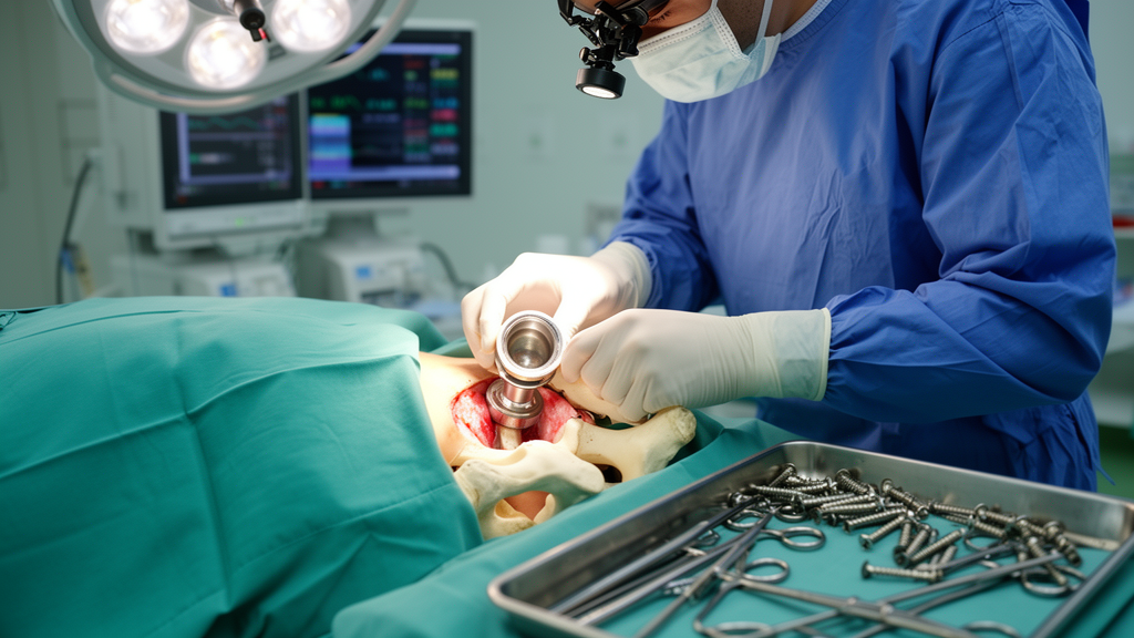
Pelvic discontinuity—a complete separation between the superior and inferior halves of the pelvis—is one of the most feared complications in revision total hip arthroplasty. When the acetabulum loses its structural integrity, conventional reconstruction techniques become unreliable, and surgeons are left with few durable options.
The study, published in Archives of Orthopaedic and Trauma Surgery, included 19 patients with intraoperatively confirmed pelvic discontinuity who underwent revision hip arthroplasty between April 2014 and April 2024. All patients had Paprosky type 3B defects, the most severe category of acetabular bone loss, characterized by extensive superior and medial bone loss with disruption of both the anterior and posterior columns. The researchers divided the patients into three groups according to the reconstruction technique used and followed them for a minimum of two years, with a mean follow-up of 51.3 months.
Group 1, comprising eight patients, received an anti-protrusio cage alone—the traditional approach in which a metal reinforcement cage bridges the discontinuity but provides only mechanical fixation. Group 2, with six patients, underwent anterior intrapelvic quadrilateral surface plating before cage implantation, adding a medial buttress that prevents cement intrusion and creates a continuous arch from the posterior to the anterior column. Group 3, the smallest cohort with five patients, underwent the novel technique: restoration of pelvic continuity through anterior and posterior column fixation with plates, followed by implantation of a porous-coated primary acetabular cup designed to achieve biological fixation through osseointegration rather than relying solely on mechanical support.
The most striking result concerned aseptic loosening, which differed significantly among the three groups (P = 0.039). Half of the cage-only patients—four of eight—developed aseptic loosening, whereas no patient in either the plate-plus-cage group or the bicolumnar fixation group experienced this complication. Reoperation rates were also markedly higher in the cage-only group, at 75 percent, compared with 16.7 percent in the plating group and 20 percent in the bicolumnar group, although this difference did not reach statistical significance (P = 0.074).
Kaplan–Meier survival analysis, with reoperation for any cause counted as an event, showed a trend toward poorer reoperation-free survival in the cage-only group, which demonstrated a progressive decline in survivorship over time. Both the plate-plus-cage group and the bicolumnar fixation group maintained 80 percent reoperation-free survival, each following a single reoperation event. However, the differences among the groups were not statistically significant on log-rank testing (P = 0.237), a reflection of the small cohort size and limited number of events.
The surgical technique behind the novel approach is technically demanding. It begins with the patient supine, using an anterior intrapelvic approach—related to the modified Stoppa approach—to expose the quadrilateral surface and anterior column, where a contoured locking plate is applied under fluoroscopic guidance. The patient is then repositioned in the lateral decubitus position, and through a posterolateral approach, the posterior column is stabilized with a reconstruction plate. Only after this bicolumnar fixation restores sufficient structural stability is the acetabulum prepared and a porous-coated primary cup implanted with supplementary screw fixation. In all three study groups, a highly cross-linked polyethylene liner was used as the bearing insert.
The rationale for using a standard primary cup rather than a cage or a complex custom implant rests on biology and economics. When bicolumnar fixation creates a stable mechanical environment, the porous-coated cup can achieve biological fixation through osseointegration, potentially improving long-term implant survival compared with cage-based constructs that depend entirely on mechanical fixation in compromised host bone. The authors also note a potential economic advantage: the technique uses widely available standard fixation implants and primary porous-coated acetabular components, avoiding the high cost and production delays associated with patient-specific triflange implants. Despite the additional fixation procedures, operative times remained comparable across the three groups.
The findings align with prior literature cited by the authors. Paprosky and colleagues previously reported loosening rates of 44 percent and rerevision rates of 31 percent with cage-only constructs, while a meta-analysis by Szczepański and colleagues found that anti-protrusio cages carried the highest mechanical failure rate (25 percent) and complication rate (34 percent) compared with cup–cage and custom triflange systems. Biomechanical studies have shown that dual-column fixation reduces micromotion, increases construct stiffness, and improves performance during early rehabilitation. Notably, the study found no meaningful differences between the plating-plus-cage and bicolumnar groups, suggesting that restoring pelvic continuity itself may matter more than the specific construct used to achieve it.
The authors are careful to frame these results as preliminary. The retrospective design, small sample size, and absence of randomization introduce selection bias, since the choice of reconstruction technique was based on preoperative imaging, bone stock quality, and intraoperative assessment rather than random allocation. Functional outcome scores were not collected, in part because the patients’ mean age of 71.2 years and severely limited ambulatory capacity made standardized functional assessment less representative. The numerically shorter follow-up in the bicolumnar group—41.2 months versus 50.1 months in the cage-only group—may have underestimated late aseptic loosening, and the small cohort restricts the robustness of the survival estimates. No bone grafting was used in any patient, due to limited availability of grafting resources at the institution.
Nevertheless, the study represents the first clinical comparison of these three reconstruction strategies for pelvic discontinuity, including the novel bicolumnar technique with primary cup implantation. For a condition so rare that most published series involve similarly small cohorts, the observation that column-stabilizing strategies were associated with lower complication and reoperation rates than cage-only reconstruction carries practical weight. The authors conclude that in selected patients, restoring pelvic continuity through column stabilization—whether with anterior plating plus a cage or with full bicolumnar fixation and a porous-coated primary cup—may offer improved mechanical stability and, in the latter case, the added potential of biological fixation. Larger prospective multicenter studies will be needed to validate these findings and confirm the long-term durability of the biological fixation achieved with primary acetabular cups in this challenging patient population.
Subject of Research: Comparative outcomes of three acetabular reconstruction techniques, including bicolumnar fixation with a primary porous-coated cup, for pelvic discontinuity in revision hip arthroplasty
Article Title: Novel bicolumnar fixation with primary cup implantation for pelvic discontinuity in revision hip arthroplasty: a comparative clinical study
Article References: Abul, M. S., Ağır, M., Yılmaz, H., Hekim, Ö., Sevim, Ö. F., & Tuncay, İ. (2026). Novel bicolumnar fixation with primary cup implantation for pelvic discontinuity in revision hip arthroplasty: a comparative clinical study. Archives of Orthopaedic and Trauma Surgery, 146(1), Article 385. https://doi.org/10.1007/s00402-026-06424-y
Image Credits: AI Generated
DOI: 10.1007/s00402-026-06424-y
Keywords: pelvic discontinuity, revision hip arthroplasty, acetabular reconstruction, anti-protrusio cage, bicolumnar fixation, porous-coated acetabular cup, Paprosky type 3B, aseptic loosening, quadrilateral surface plating, osseointegration, reoperation-free survival, orthopedic surgery
News Source: Ophelia Keating. (October 29, 2026). Bicolumnar Fixation With Primary Cup Shows Promise in Rare Hip Revision Challenge. Scienmag.