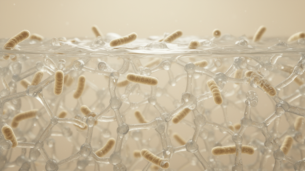
A strain of Lactiplantibacillus plantarum can turn alginate into a gel without receiving a conventional crosslinking agent, according to a laboratory study that characterized the phenomenon in culture media containing sodium alginate. Researchers at Tohoku University found that reproducible gelation occurred when 1% weight-per-volume sodium alginate was cultivated with the bacterium for 24 hours at 37 °C and then incubated at 4 °C for one hour. Under those conditions, the culture changed from a liquid into a homogeneous, extrudable material that retained its shape when the tube was inverted. The work establishes an initial biological route to alginate gel formation, but it does not identify the specific extracellular molecule or molecules responsible or demonstrate performance in a clinical or commercial application.
The study began from an observation made during earlier work on cultivating L. plantarum in calcium alginate materials. The researchers noticed that bacteria grown in MRS broth containing 1% sodium alginate at 25 °C became a gel after subsequent storage at 4 °C, even though no external crosslinker had been added. Fresh alginate solution exposed to the same low temperature remained fluid. That contrast suggested that the bacteria were altering the polymer environment during growth, but it did not establish whether the cells, their metabolites or some combination of the two were responsible. The present experiment was therefore designed as an initial characterization of a previously unreported biological gelation process rather than as a fully optimized material formulation.
To test the requirements for gel formation, the team varied sodium alginate concentration, bacterial cultivation time and post-cultivation temperature. Gelation was judged macroscopically with a standardized calcium alginate bead: in a liquid sample the bead sank, whereas a newly formed gel matrix supported it near the surface. This indicator was not used to measure gel strength or other mechanical properties. At an alginate concentration of 0.5%, gel formation occurred only under limited conditions. A concentration of 1% consistently produced stable gels after the bacteria had been cultivated, while 2% supported gelation across a broader set of conditions. The results indicate that bacterial growth and a sufficient density of polymer chains are both required for the process. The standard preparation used for subsequent experiments was 1% alginate, 24 hours of bacterial cultivation and one hour at 4 °C.
Fractionating the culture provided the central mechanistic clue. The researchers prepared whole bacterial cultures, washed bacterial cells resuspended in water, and cell-free supernatants obtained by centrifuging and filtering the culture. Whole cultures and cell-free supernatants induced visible gel formation after low-temperature incubation, whereas the resuspended bacterial cells did not. This separation points to extracellular products of L. plantarum, rather than the physical presence of intact cells, as the active fraction under the tested conditions. The finding does not yet identify the relevant product, and the authors emphasize that the supernatant could contain several candidate contributors, including organic acids, exopolysaccharides, proteins, extracellular DNA and other metabolites.
Acification was the next focus of the investigation. L. plantarum primarily produces lactic acid during carbohydrate fermentation, and the bacterial cultures used for gelation reached a pH of approximately 4.3. The researchers separately acidified alginate-containing medium with lactic acid, a mixture of lactic and acetic acids, or hydrochloric acid, bringing each solution to the same approximate pH rather than matching a predetermined acid concentration. All of these chemically acidified samples underwent visible gelation after incubation at 4 °C. The result shows that a sufficiently acidic environment can initiate gel formation under the experimental conditions; it does not establish that lactic acid alone is responsible. Lowering the pH would be expected to protonate alginate carboxylate groups, reducing electrostatic repulsion between negatively charged chains and promoting intermolecular association.
Scanning electron microscopy nevertheless revealed a difference between simple acidification and biological gelation. Alginate cultured with L. plantarum had a dense appearance before cold incubation and developed a porous three-dimensional network afterward. A similar network appeared in gels formed by cell-free supernatant. By contrast, the gels produced with defined acids had comparatively compact morphologies. The authors therefore interpret acidification as a major trigger of gel formation, while additional extracellular products appear to influence the extent of association and the architecture of the resulting network. The molecular basis of the need for low-temperature incubation remains unresolved. One possibility discussed by the researchers is that acidification creates a state permissive for gelation, after which cooling promotes physical rearrangement or association among protonated alginate chains.
The newly formed material also retained useful processing characteristics. When extruded into distilled water, it formed a continuous filament before gradually dispersing. After being written or shaped, it could be sprayed with 1.5% calcium chloride and held for 20 minutes to produce a mechanically stabilized construct that retained its three-dimensional geometry. The researchers demonstrated extrusion, direct writing of patterned letters, and spreading onto skin; the skin coating could be rinsed away with running water. Compared with non-cultured alginate subjected to the same secondary calcium treatment, the bacteria-induced gel contracted less. These findings indicate that the spontaneous gelation changes how the polymer subsequently responds to calcium. They are qualitative processing observations, however, and the study did not systematically measure rheology, swelling, degradation, long-term mechanical stability or tensile strength.
Bacterial survival was assessed after the gels were stored for up to seven days at 4 °C, 25 °C or 37 °C. At 4 °C, L. plantarum remained highly viable throughout the storage period, with only a slight reduction in colony-forming units after seven days. Viability declined more gradually at 25 °C and more markedly at 37 °C. Indicator strips embedded in the gel changed little in color, suggesting that the acidic microenvironment created during cultivation remained broadly stable under the tested conditions. The authors interpret the temperature-dependent losses as possible consequences of continued metabolism, nutrient depletion and metabolic stress at warmer temperatures, while refrigeration slows those processes. The experiment establishes refrigerated viability for the tested period and conditions, not a validated shelf life or evidence that the gel can preserve bacteria during transport or long-term distribution.
The findings open several avenues for biofabrication while leaving important limitations. Because gel formation occurred while bacteria were present and viable, the approach could eventually inform live probiotic bioinks, living materials and gel systems that combine bacterial activity with a polysaccharide matrix. The spreadable material may also serve as a carrier for locally retained bacterial products, and the active gelation capacity of the cell-free supernatant suggests possible postbiotic gels that contain bacterial metabolites without living cells. These are proposed directions rather than demonstrated applications: the study did not test wound healing, tissue repair, drug delivery, printing performance in a controlled bioink system, or therapeutic efficacy. It examined only L. plantarum, used a complex MRS medium, and reported modest biological variation in final pH and extracellular composition. Quantitative material characterization was also limited, while the active gelation factor, the basis of temperature dependence, and whether other bacterial species produce similar or distinctive structures remain open questions. The study therefore provides a reproducible demonstration of bacteria-directed alginate gelation and a foundation for subsequent biochemical fractionation, controlled reconstruction and comparative testing.
Subject of Research: Bacteria-induced spontaneous gelation of sodium alginate using extracellular products of Lactiplantibacillus plantarum
Article Title: Biologically induced spontaneous alginate gelation mediated by extracellular products of Lactiplantibacillus plantarum
Article References: Gong, W., Cisterna-Vergara, S., Kanazawa, M., Villena, J., Nishiyama, K., & Kitazawa, H. (2026). Biologically induced spontaneous alginate gelation mediated by extracellular products of Lactiplantibacillus plantarum. Frontiers in Bioengineering and Biotechnology, 14, Article 1955981. https://doi.org/10.3389/fbioe.2026.1955981
Image Credits: AI Generated
DOI: 10.3389/fbioe.2026.1955981
Keywords: alginate, bacterial gelation, Lactiplantibacillus plantarum, extracellular products, biofabrication, living biomaterials, hydrogels, acidification, calcium crosslinking, bacterial viability, Biologically, induced
News Source: Denise Maddox. (October 20, 2026). Bacterial products trigger spontaneous alginate gel formation without added crosslinkers. Scienmag.