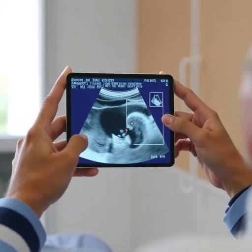
In a groundbreaking advancement that promises to revolutionize medical ultrasound imaging, researchers at the Massachusetts Institute of Technology (MIT) have developed an innovative technique that transforms traditional two-dimensional scans into immersive, real-time three-dimensional visualizations using augmented reality (AR). This novel approach, unveiled in a study published today in Communications Engineering, offers unprecedented clarity and precision in visualizing internal anatomical structures, fundamentally enhancing both medical training and clinical procedures.
Ultrasound imaging, a ubiquitous modality in medical diagnostics, traditionally relies on interpreting a series of two-dimensional slices to mentally reconstruct a three-dimensional representation of tissues and organs. This reliance on mental spatial reasoning presents a steep cognitive challenge, especially for novice users, often leading to inaccuracies in diagnosis and procedural guidance. The new MIT method leverages advances in 3D ultrasound capture combined with augmented reality display technology to make this process intuitive and accurate.
The core innovation lies in integrating a specialized ultrasound probe with a real-time volumetric imaging system. This compact probe, smaller than a standard deck of cards, employs a chirped data acquisition (cDAQ) system and features an ultrasound array arranged in an open square formation. This configuration efficiently captures volumetric data from below the probe, enabling affordable, low-power three-dimensional imaging. Unlike conventional 3D ultrasound systems, which are costly and complex, this approach democratizes access by simplifying hardware requirements without compromising image fidelity.
Captured ultrasound voxel data is instantly processed and rendered using the Unreal Engine, a powerful 3D graphics platform normally utilized in gaming and virtual simulations. This conversion transforms raw ultrasound signals into precise, positionally accurate 3D models of scanned tissues. By donning AR or virtual reality (VR) headsets, users are presented with a vivid “X-ray vision” view, overlaying these models directly on the physical body or object under examination. This spatial alignment allows clinicians to manipulate their perspective naturally, rotating or zooming to grasp complex anatomical relationships effortlessly.
The team named their system AR-VIU, standing for Augmented Real-time Volumetric Imaging in Ultrasound. To rigorously assess its clinical and educational impact, they recruited 18 participants split evenly between experienced sonographers and ultrasound novices. Subjects performed tasks of identifying embedded objects inside opaque gelatin containers and marking precise target sites on tissue-mimicking phantoms, emulating clinical needle placement for biopsies.
Results decisively demonstrated that AR-VIU narrowed performance gaps between experts and novices. Novices using AR-enhanced 3D imaging nearly matched the accuracy of trained experts, a substantial improvement compared to conventional 2D ultrasound methods. This suggests that superimposing volumetric data with real-world spatial cues profoundly reduces the cognitive load typically associated with interpreting ultrasound images, making the technology accessible to a wider range of healthcare providers with less training required.
Experienced professionals, while favoring the familiarity of 2D imaging, acknowledged the considerable advantages AR-VIU offers in specific challenging contexts such as cardiac wall motion analysis during echocardiography or precise needle guidance in interventional procedures. The intuitive nature of real-time 3D feedback holds promise for reducing procedure times and increasing confidence in clinically critical moments.
From a technical perspective, the AR-VIU technology capitalizes on efficient compression and streaming protocols to transmit volumetric data seamlessly to the AR display without latency. This ensures live interaction with the imaging field, an essential factor for dynamic clinical applications. Future development efforts center on enhancing image resolution further, validating diagnostic accuracy in diverse clinical environments, and integrating the system into conventional hospital workflows.
This breakthrough demonstrates the transformative potential when cutting-edge imaging physics meets immersive computing. By effectively bridging the gap between complex volumetric data and human perceptual capabilities, the MIT team has opened a new horizon for non-invasive diagnostics and interventional guidance. Their work exemplifies how emerging technologies can dismantle long-standing barriers in medical practice, ultimately improving patient outcomes and training efficiency worldwide.
In addition to the clinical implications, this technology may catalyze innovation in medical education. Training ultrasound technicians traditionally demands extensive practice and mentoring to overcome the challenging visual-spatial interpretation. AR-VIU’s immersive display accelerates learners’ grasp of anatomy and probe manipulation, reducing time to proficiency and fostering deeper understanding.
The research team acknowledges support from the MIT Media Lab Consortium, the U.S. National Science Foundation, and prestigious fellowships contributing to this project. Lead authors graduate students Jason Hou and Shrihari Viswanath, alongside collaborators including senior researchers and summer interns, have pioneered this effort, setting a new benchmark for future ultrasound imaging modalities.
Looking forward, the fusion of real-time volumetric ultrasound with augmented reality could extend beyond typical clinical scenarios. Potential applications range from guiding robotic surgery to enabling remote consultations with enhanced visual context. As AR headsets become more compact and affordable, such systems may see widespread adoption across diverse healthcare settings, from rural clinics to specialized centers.
In summary, MIT’s AR-VIU system heralds a new age in ultrasound imaging, marking a significant step toward fully immersive, easy-to-interpret medical imaging that bridges the gap between novice and expert performance. By transforming abstract 2D slices into intuitive 3D experiences, it empowers a broader spectrum of healthcare professionals to provide faster, safer, and more accurate diagnostic and therapeutic care.
Subject of Research: People
Article Title: Real-time 3D ultrasound in augmented reality accelerates training and narrows novice–expert performance gaps
News Publication Date: 10-Jun-2026
Web References: 10.1038/s44172-026-00692-7
Image Credits: MIT
Keywords: Health and medicine, Physical sciences, Ultrasound, Ultrasonics, Imaging, Medical imaging, Cancer, Research methods
Tags: 3D anatomical structure visualization3D ultrasound visualization technologyaugmented reality for medical trainingaugmented reality in medical ultrasoundclinical ultrasound procedure enhancementcognitive challenges in ultrasound interpretationimproving ultrasound diagnostic accuracylow-power 3D ultrasound devicesmedical imaging augmented reality applicationsMIT ultrasound innovationreal-time volumetric ultrasound imagingultrasound probe with chirped data acquisition