Credit: (left) Andrey Ryabov, LMU Munich; (right) Mikhail Volkov, University of Konstanz
Electron microscopes provide deep insight into the smallest details of matter and can reveal, for example, the atomic configuration of materials, the structure of proteins or the shape of virus particles. However, most materials in nature are not static and rather interact, move and reshape all the time. One of the most common phenomena is the interaction between light and matter, which is ubiquitous in plants as well as in optical components, solar cells, displays or lasers. These interactions – which are defined by electrons being moved around by the field cycles of a light wave – happen at ultrafast time scales of femtoseconds (10-15 seconds) or even attoseconds (10-18 seconds, a billionth of a billionth of a second). While ultrafast electron microscopy can provide some insight into femtosecond processes, it has not been possible, until now, to visualize the reaction dynamics of light and matter occurring at attosecond speeds.
Now, a team of physicists from the University of Konstanz and Ludwig-Maximilians-Universität München have succeeded in combining a transmission electron microscope with a continuous-wave laser to create a prototypical attosecond electron microscope (A-TEM). The results are reported in the latest issue of Science Advances.
Modulating the electron beam
“Basic phenomena in optics, nanophotonics or metamaterials happen at attosecond times, shorter than a cycle of light”, explains Professor Peter Baum, lead author on the study and head of the Light and Matter research group at University of Konstanz’s Department of Physics. “To be able to visualize ultrafast interactions between light and matter requires a time resolution below the oscillation period of light”. Conventional transmission electron microscopes use a continuous electron beam to illuminate a specimen and create an image. To achieve attosecond time resolution, the team led by Baum uses the rapid oscillations of a continuous-wave laser to modulate the electron beam inside the microscope in time.
Ultra-short electron pulses
Key to their experimental approach is a thin membrane which the researchers use to break the symmetry of the optical cycles of the laser wave. This causes the electrons to accelerate and decelerate in rapid succession. “As a result, the electron beam inside the electron microscope is transformed into a series of ultrashort electron pulses, shorter than half an optical cycle of the laser light”, says first author Andrey Ryabov, a postdoctoral researcher on the study. Another laser wave, which is split from the first one, is used to excite an optical phenomenon in a specimen of interest. The ultrashort electron pulses then probe the sample and its reaction to the laser light. By scanning the optical delay between the two laser waves, the researchers are then able to obtain attosecond resolution footage of the electromagnetic dynamics inside the specimen.
Simple modifications, large impact
“The main advantage of our method is that we are able to use the available continuous electron beam inside the electron microscope rather than having to modify the electron source. This means that we have a million times more electrons per second, basically the full brightness of the source, which is key to any practical applications”, continues Ryabov. Another advantage is that the necessary technical modifications are rather simple and do not require electron gun modifications.
As a result, it is now possible to achieve attosecond resolution in a whole range of space-time imaging techniques such as time-resolved holography, waveform electron microscopy or laser-assisted electron spectroscopy, amongst others. In the long term, attosecond electron microscopy may help to uncover the atomistic origins of light-matter interactions in complex materials and biological substances.
###
Facts:
- Ultrafast imaging breakthrough: Physicists from the University of Konstanz and Ludwig-Maximilians-Universität München in Germany achieve attosecond time resolution in a transmission electron microscope by combining it with a continuous-wave laser.
- Research team led by Professor Peter Baum (University of Konstanz) modify a transmission electron microscope to create time-resolved images of light-matter interactions at attosecond speeds (10-18 seconds).
- Potential boost for a range of imaging techniques and the further exploration of the atomistic origins of light-matter interactions.
- Original publication: A. Ryabov, J. W. Thurner, D. Nabben, M. V. Tsarev, P. Baum, Attosecond metrology in a continuous-beam transmission electron microscope, Science Advances, 11 November 2020. DOI: 10.1126/sciadv.abb1393. URL: https:/
/ (will become active after the embargo has lifted).advances. sciencemag. org/ lookup/ doi/ 10. 1126/ sciadv. abb1393 - Embargoed: Not for release until Wednesday, 11 November 2020, at 14:00 US Eastern Time.
Note to editors:
You can download a photo here: https:/
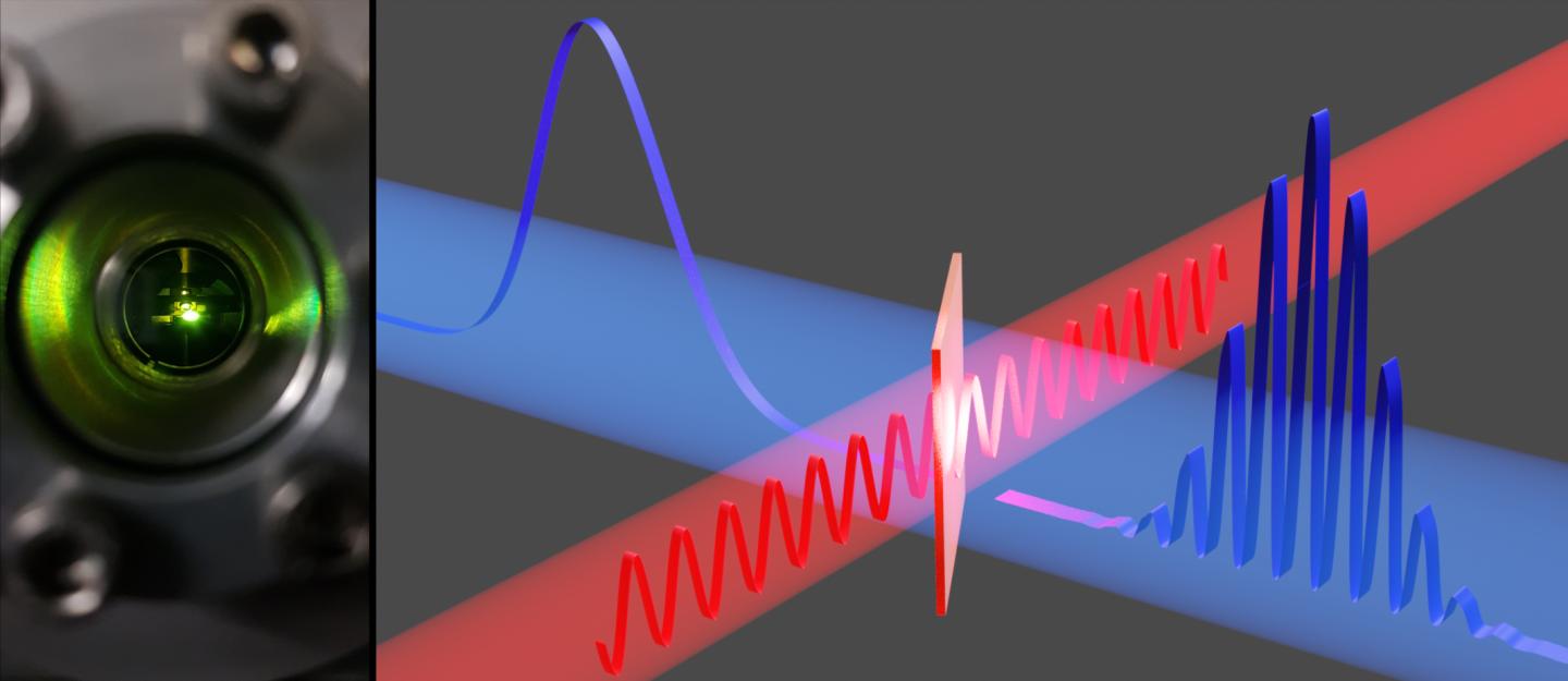
Caption: (left) A glance inside an attosecond transmission electron microscope. (right) A continuous-wave laser (red) intersects with an electron beam (blue) at a membrane. The laser light bunches the electrons (blue wavelet) into an attosecond pulse train (modulated wavelet).
Image credit: (left) Andrey Ryabov, LMU Munich; (right) Mikhail Volkov, University of Konstanz
Contact:
University of Konstanz
Communications and Marketing
Phone: +49 7531 88-3603
Email: [email protected]
uni.kn/en
Media Contact
Julia Wandt
[email protected]
Related Journal Article
http://dx.


