In the advancing realm of medical imaging, the segmentation of lymph nodes (LNs) within uterine MRI scans presents a critical challenge, largely due to the subtle and unclear boundaries, varying shapes, and size diversity of lymphatic structures. A groundbreaking study now introduces an innovative deep learning methodology that leverages the synergy of bimodal magnetic resonance imaging (MRI) and a novel attention-enhanced residual U-Net architecture, fundamentally refining the precision of LN detection and segmentation.
Lymph nodes are vital diagnostic markers in many gynecological diseases, including uterine cancers. Their precise identification in MRI scans is indispensable for accurate staging and treatment planning. However, the inherent difficulty posed by lymph nodes’ indistinct borders and similarity with adjacent tissue has historically impeded reliable automation efforts. Recognizing this obstacle, researchers combined two MRI modalities—T2-weighted imaging (T2WI) and diffusion-weighted imaging (DWI)—to complement each other’s strengths, thereby enriching the input data with valuable anatomical and pathological information.
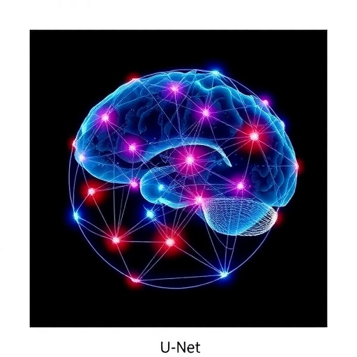
This dual-modal imaging approach feeds into a meticulously designed deep neural network: the Efficient Residual U-Net (ERU-Net). This architecture transcends traditional U-Net models by incorporating an efficient channel attention (ECA) mechanism and a residual network framework within its encoder-decoder structure. The ECA module dynamically recalibrates feature maps by emphasizing salient channels, enabling the network to better discriminate lymph nodes from surrounding tissues. Simultaneously, the residual connections facilitate a deeper network capable of learning complex hierarchical features without degradation, ensuring robust segmentation outputs.
.adsslot_1OLiUCVzbX{ width:728px !important; height:90px !important; }
@media (max-width:1199px) { .adsslot_1OLiUCVzbX{ width:468px !important; height:60px !important; } }
@media (max-width:767px) { .adsslot_1OLiUCVzbX{ width:320px !important; height:50px !important; } }
ADVERTISEMENT
Manual annotations were painstakingly generated by two expert radiologists, ensuring that the ground truth labels upheld the highest standards of clinical accuracy. The dual-modality images were pixel-wise fused before being presented to the ERU-Net, allowing simultaneous exploitation of the nuanced contrast variations from T2WI and the diffusion metrics embedded in DWI scans. This innovative pre-processing step critically enhanced the contextual richness available to the neural network.
Evaluation metrics underscored the significant performance leap achieved by this approach. The ERU-Net attained a mean intersection-over-union (mIoU) of 0.83, a figure that represents a remarkably precise overlap between automated segmentation and expert delineations. Furthermore, the network’s average pixel accuracy reached 91%, while precision and recall metrics stood at 0.90 and 0.91 respectively—indicators of balanced sensitivity and specificity critical for clinical utility.
Comparative analyses revealed the superiority of ERU-Net over existing segmentation networks in the task of uterine lymph node delineation. Conventional U-Net variants and other state-of-the-art models suffered from less consistent boundary localization or failed to simultaneously optimize precision and recall. The integration of efficient channel attention and residual structures synergistically addressed these limitations by enhancing feature representation and mitigating vanishing gradient issues common in deep architectures.
The implications of this technology transcend mere algorithmic achievement. Accurate and automated segmentation tools streamline radiological workflows, reduce diagnostic subjectivity, and expedite therapeutic decision-making. For patients, this translates to earlier and more precise interventions, potentially improving prognoses in diseases where lymph node status is a pivotal factor. From a healthcare system perspective, the reduction in manual annotation requirements and interpretation time heralds a cost-effective future for imaging diagnostics.
Moreover, the methodology’s embrace of bimodal imaging points toward a larger paradigm shift where multimodal data fusion becomes standard in medical AI. By judiciously combining complementary imaging techniques, machine learning systems can extract richer pathology signatures, facilitating earlier detection and nuanced disease characterization previously unattainable with single-modality inputs.
The study also highlights opportunities for further technology refinement. Incorporating additional MRI sequences, integrating temporal dynamics from longitudinal imaging, or adapting the ERU-Net framework for other anatomical sites represent promising directions. Beyond segmentation, the extracted features from attention-enhanced residual networks could feed into predictive models anticipating disease progression, therapeutic response, or patient outcomes.
While the current dataset is notable, expanding the scale and diversity—across demographics and imaging hardware—will be essential to validate the robustness and broader applicability of the ERU-Net. Additionally, integrating explainability modules into the network could enhance clinician trust by visually demonstrating decision-making pathways within the segmentation process, aligning with the pressing demand for transparent AI in medicine.
In conclusion, the introduction of the Attention-enhanced residual U-Net marks a transformative milestone in MRI-based lymph node segmentation. By fusing dual-modal imaging data and harnessing innovative attention mechanisms within a residual deep learning framework, this approach achieves unprecedented segmentation accuracy. This breakthrough not only elevates the technical frontier of automated medical image analysis but also promises tangible clinical benefits, setting a blueprint for future AI-driven diagnostic tools.
The research, published in BioMedical Engineering OnLine, exemplifies the power of interdisciplinary collaboration between radiology and artificial intelligence, reinforcing the role of cutting-edge computational methods in reshaping modern healthcare. As imaging datasets grow and computational resources evolve, such sophisticated models hold the key to unlocking new horizons in personalized medicine.
The ERU-Net’s success story underscores the broader narrative of AI’s integration into clinical practice: harnessing complexity with simplicity, translating raw multimodal data into actionable insights, and ultimately enhancing patient care. This visionary work inspires ongoing efforts to deepen machine understanding of human anatomy and pathology, steering the future of diagnostic radiology toward unprecedented precision and reliability.
Subject of Research: Lymph node segmentation in uterine MRI images using an attention-enhanced residual U-Net architecture with bimodal imaging data.
Article Title: Attention-enhanced residual U-Net: lymph node segmentation method with bimodal MRI images
Article References:
Qiu, J., Chen, C., Li, M. et al. Attention-enhanced residual U-Net: lymph node segmentation method with bimodal MRI images.
BioMed Eng OnLine 24, 67 (2025). https://doi.org/10.1186/s12938-025-01400-w
Image Credits: AI Generated
DOI: https://doi.org/10.1186/s12938-025-01400-w
Tags: attention-enhanced U-Net architectureautomation in medical imagingdeep learning in medical imagingdual-modal MRI imaging techniquesefficient channel attention mechanismgynecological disease stagingimproving segmentation accuracy in MRIlymph node segmentation in MRIlymphatic structure detection challengesresidual U-Net for medical applicationsT2-weighted and diffusion-weighted MRIuterine cancer diagnosis

