Astigmatism affecting nearly three-quarters of examined eyes, a tiger-striped retinal appearance in half, and upward-slanting eyelid fissures in almost every child tested: those are the headline numbers from a new cross-sectional study of 48 Egyptian children with trisomy 21, the chromosomal condition better known as Down syndrome. The research, conducted at Cairo University Specialized Pediatric Hospital and published in the Egyptian Pediatric Association Gazette, systematically catalogued eye abnormalities in patients aged one to nine years and then looked for statistical links between those ocular findings and the children’s systemic features, including congenital heart disease and thyroid status.
The study enrolled children with cytogenetically confirmed trisomy 21 from the hospital’s Medical Genetics and Pediatric Ophthalmology Clinics between March 2021 and December 2023. It was a non-random, convenience sample, a design choice the authors acknowledge may limit how broadly the results apply. Children with a history of ocular trauma or previous eye surgery were excluded. The final cohort comprised 29 boys (60.4 percent) and 19 girls (39.6 percent), with a mean age of 6.2 years and a range spanning one to nine years. Mean maternal age at delivery was 39.5 years and mean paternal age 45.1 years, and consanguinity was recorded in 22.9 percent of families.
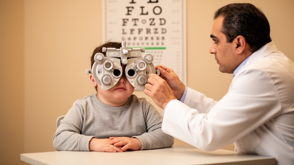
Every child received a comprehensive ophthalmologic examination performed by a pediatric ophthalmologist. The workup covered external and eyelid assessment, including epicanthal folds, ptosis, blepharitis and the slanting of the palpebral fissures; visual acuity testing with a chart projector when the child could cooperate; ocular motility evaluation using cover-uncover and prism bar methods; anterior segment inspection with a handheld slit lamp; and posterior segment examination by indirect ophthalmoscopy with a +20 D lens to assess the vitreous, retina, retinal vessels and optic nerve. Intraocular pressure was measured with a Perkins applanation tonometer, and cycloplegic refraction was obtained with an autorefractometer and retinoscopy. Uncooperative children were sedated for retinoscopy and slit-lamp examination using oral chloral hydrate syrup, with topical benoxinate anesthesia applied.
The refractive error results were striking. Astigmatism was the most frequent abnormality, present in 71.9 percent of eyes, or 69 eyes across the cohort. Hyperopia appeared in 40.6 percent (39 eyes) and myopia in 36.4 percent (35 eyes). The researchers defined ametropia, meaning hyperopia, myopia or astigmatism, as a refractive error of 0.75 diopters or more. In the discussion, the authors note that their astigmatism figure agrees with prior reports exceeding 70 percent, and that the hyperopia and myopia rates fall within ranges published elsewhere, including a Colombian study that found hyperopia in 41.8 percent and myopia in 29.9 percent of trisomy 21 patients. The authors suggest the high prevalence of both spherical refractive errors supports the hypothesis that emmetropization, the developmental process by which the eye’s optical components normally come into focus, fails in Down syndrome.
Posterior segment findings added another layer. Tigroid retina, a pigmented mottled appearance of the fundus, was found in 51 percent of eyes (49 eyes), making it the most prevalent posterior segment abnormality in this cohort and exceeding the 36 percent figure the authors cite from a literature review. Tortuous retinal vessels appeared in 8.3 percent of eyes, peripapillary atrophy in 6.3 percent, pale disc in 5.2 percent, pigmentary retinal abnormalities in 4.2 percent, and hyperemic disc in 27.1 percent. Mean intraocular pressure across the group was 13.19 millimeters of mercury, with individual readings ranging from 7 to 20 millimeters of mercury.
Anterior segment and eyelid findings were equally prominent. Upward slanting palpebral fissures were recorded in 93.8 percent of eyes, blepharitis in 20.8 percent, and conjunctivitis in 14.6 percent. Cataract was present in 5.2 percent of eyes and glaucoma in just one eye, or 1 percent of the studied group. Notably, no corneal anomalies were observed, and keratoconus was absent. The authors attribute the lack of keratoconus to the young age of the cohort, since that cone-shaped corneal thinning disorder typically develops in adolescence and early adulthood, beyond the age range studied here.
Among the muscles and adnexal structures, strabismus was identified in 25 percent of patients, predominantly esotropia, the inward-turning form, which accounted for 12.5 percent. Nystagmus, the rhythmic involuntary movement of the eyes, was observed in 14.6 percent of patients. Nasolacrimal duct obstruction, which blocks tear drainage, was found in 2.1 percent. The authors place these figures in international context: literature reviews cited in the paper report strabismus in roughly a third of trisomy 21 patients, esotropia in about 28 percent, and nystagmus in about 16 percent, while glaucoma prevalence across published studies ranges from zero to 6.7 percent.
The systemic side of the analysis revealed substantial comorbidity. Congenital heart disease was present in 45.8 percent of the children, or 22 patients, with patent foramen ovale the most common lesion at 18.8 percent. Hypothyroidism affected three patients, 6.3 percent of the group. Skeletal features were common as well: sandal gap, a widened space between the first two toes, appeared in 47.9 percent, clinodactyly, the incurving of a finger, in 39.6 percent, and a simian crease in 37.5 percent. Underweight status was present in 22.9 percent and stunted growth in 8.3 percent, while blood pressure and heart rate were within normal limits for all participants.
Perhaps the most intriguing results were the statistical correlations between ocular and systemic features, assessed with Fisher’s exact test at a significance threshold of p below 0.05. Clinodactyly correlated with optic nerve hypoplasia, myopia, myopic retina, strabismus and nystagmus. Sandal gap correlated with blepharitis, iris thinning, pigmentary retinal abnormalities, tortuous retinal vessels and strabismus. Congenital heart disease showed associations with blepharitis, cataract, tortuous retinal vessels and hyperemic optic disc. The authors propose a possible mechanism for the cardiac link: vascular anomalies secondary to congenital heart disease may lead to retinal and optic nerve changes, a hypothesis they connect to prior literature on cardiocular associations.
The authors are careful about the study’s boundaries. Beyond the small sample, single-center design and convenience sampling, the cross-sectional nature prevented any assessment of how findings progress over time. The young age range may have underestimated age-related conditions such as keratoconus. Some systemic investigations were performed only according to availability, and the paper mixes per-eye and per-patient reporting, which complicates direct comparison between different findings. Those caveats matter for interpretation, but they do not erase the central signal: ocular abnormalities were highly prevalent in this cohort, refractive errors led the list, and several eye findings tracked with systemic features.
The practical implication the authors draw is straightforward. They conclude that ocular abnormalities are highly prevalent among children with trisomy 21 compared with the general population, and that comprehensive ophthalmologic evaluation should begin early in life. Early detection of serious conditions such as glaucoma and cataract is described as essential for improving visual prognosis, while refractive errors, the most common finding, can be addressed through regular screening and timely correction. The observed correlations between ocular and systemic manifestations, they argue, reinforce the case for a multidisciplinary approach in which pediatric ophthalmology, cardiology, endocrinology and genetics work together in the care of children with Down syndrome.
Subject of Research: Ophthalmic manifestations and systemic correlations in pediatric trisomy 21 at a Cairo hospital
Article Title: Ophthalmic manifestations of trisomy 21 syndrome in the pediatric age group: a cross-sectional study from Cairo University Specialized Pediatric Hospital
Article References: Abdelmohsen, E. W., Abdel Meguid, I. E., Kassem, R. R., Amer, S. T., El Gindy, H. A., Shawky, S. A., & Mohamed, R. (2026). Ophthalmic manifestations of trisomy 21 syndrome in the pediatric age group: a cross-sectional study from Cairo University Specialized Pediatric Hospital. Egyptian Pediatric Association Gazette, 74(1), Article 118. https://doi.org/10.1186/s43054-026-00620-6
Image Credits: AI Generated
DOI: 10.1186/s43054-026-00620-6
Keywords: Down syndrome, trisomy 21, pediatric ophthalmology, astigmatism, strabismus, congenital heart disease, cross-sectional study, refractive errors, Egypt, retinal abnormalities, glaucoma, nystagmus
News Source: Harold Sullivan. (October 29, 2026). Astigmatism Tops Eye Findings in Egyptian Children with Down Syndrome Study. Scienmag.