DNA double-strand breaks are among the most dangerous forms of genetic damage a cell can experience. When both strands of the DNA double helix are severed, the chromosome can lose genetic information, rearrange, or break apart entirely. A new review in Applied Microbiology and Biotechnology examines how an unusual archaeal enzyme called NurA helps cells begin repairing these lesions, and why the protein may offer clues about the ancient origins of DNA repair systems shared across the tree of life.
The review, published on 26 August 2026 by Xinyan Zou, Botao Zhang, Binxian Gu, Yong Gong, Yanchao Bai and Likui Zhang, focuses on NurA as a central component of a compact DNA end-resection machine. In archaeal cells, NurA works together with the HerA helicase and the Mre11-Rad50 complex. Their combined task is to process the broken ends of DNA and generate single-stranded DNA tails with a free 3′ end. These tails are essential substrates for homologous recombination, a repair pathway that uses an intact DNA molecule as a template to restore missing genetic information.
The first challenge after a double-strand break is not simply joining the broken ends. For homologous recombination to begin, the cell must carefully reshape the damaged DNA. One strand is progressively removed from each broken end, exposing the complementary strand as single-stranded DNA. This directional process, known as end resection, creates a molecular platform on which recombination proteins can assemble. The resulting 3′ single-stranded tails are particularly important because they can search for and pair with a matching sequence in an undamaged chromosome. NurA appears to perform a major part of the enzymatic cutting required to produce these tails, while HerA helps unwind the DNA and Mre11-Rad50 contributes to the initial recognition and processing of the break.
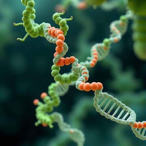
Structurally, archaeal NurA forms a toroidal dimer, producing a ring-shaped molecular assembly with a central channel. This architecture is more than a visual feature. A ring-shaped nuclease can help confine DNA within a defined catalytic environment, positioning the nucleic acid for controlled cleavage as it passes through or interacts with the channel. The dimeric arrangement also creates an extended surface for DNA binding and may help coordinate the movement of DNA with the action of the associated HerA helicase. According to the review, NurA and HerA can form a continuous channel, suggesting that DNA may be transferred directly from the helicase into the nuclease without freely diffusing into the surrounding cell.
NurA possesses two related but distinct catalytic capabilities. It can act as a 5′-to-3′ exonuclease, removing nucleotides sequentially from a DNA end, and it can also function as an endonuclease, cutting within a DNA strand rather than only at its terminus. These activities provide the enzyme with flexibility during repair. Exonucleolytic digestion can enlarge a resected region from an existing DNA end, while endonucleolytic cleavage may help initiate processing at particular DNA structures or positions. The balance between the two activities is likely to depend on the DNA substrate, the arrangement of the protein complex and the presence of partner enzymes, although the review emphasizes that important mechanistic questions remain unresolved.
The chemical engine behind NurA’s activity is a divalent metal ion. In particular, manganese ions, or Mn²⁺, are described as essential for catalysis. Like many nucleases, NurA is thought to use metal ions to organize the DNA substrate, activate water molecules and stabilize the negatively charged reaction intermediates that form when phosphodiester bonds are broken. The requirement for Mn²⁺ highlights the importance of the enzyme’s catalytic environment: without the appropriate metal cofactor, the chemical steps needed to cleave DNA cannot proceed efficiently. The review also summarizes the identification of amino acids that contribute to DNA binding and strand cleavage, helping define how NurA recognizes its substrate and positions the scissile phosphate bond for hydrolysis.
The NurA–HerA partnership is particularly significant because it links two physically different operations: DNA unwinding and DNA degradation. HerA is a helicase that uses energy from nucleotide hydrolysis to separate the two strands of the DNA duplex. NurA, positioned alongside it, can then process the exposed strand. Their continuous channel offers a possible explanation for how archaeal cells coordinate these reactions while minimizing the risk of uncontrolled DNA degradation. Rather than allowing a nuclease and helicase to operate independently, the paired system could guide DNA through a protected molecular corridor in which unwinding and resection are synchronized. The Mre11-Rad50 complex adds another layer of coordination by participating in the detection and early processing of broken chromosome ends.
This compact repair system is also important from an evolutionary perspective. NurA homologs are found across archaeal lineages and in some bacteria, but no NurA counterpart is known in eukaryotes. At the same time, eukaryotic cells possess more elaborate DNA end-resection pathways that perform related functions through different protein assemblies. The presence of NurA in archaea, together with its partnership with HerA and Mre11-Rad50, raises questions about how ancient DNA repair strategies were reorganized during the transition from prokaryotic ancestors to the first eukaryotic cells. The review presents NurA as a potentially valuable molecular marker for tracing which parts of double-strand break repair machinery were retained, replaced or lost during evolution.
Particular attention is drawn to the Asgard superphylum, a group of archaea considered especially relevant to studies of eukaryotic origins. Genomic surveys have identified widespread, previously uncharacterized NurA homologs in Asgard archaea. These proteins could reveal whether the structural and catalytic features observed in other archaeal NurA enzymes were already present in lineages related to the ancestors of eukaryotes. However, the review makes clear that Asgard-derived NurA proteins have not yet been experimentally characterized. Their biochemical activities, metal requirements, oligomeric states and interactions with HerA or Mre11-Rad50 remain open questions. Sequence similarity alone cannot establish whether these proteins possess the same dual nuclease activities or assemble into comparable DNA-processing channels.
The authors therefore identify structural biology and biochemistry as priorities for the next stage of research. Determining the three-dimensional structures of Asgard NurA proteins, ideally in complexes with DNA, HerA and metal ions, could show whether their active sites and central channels resemble those of previously studied archaeal enzymes. Complementary experiments could measure their exonuclease and endonuclease activities, define their substrate preferences and test how mutations in DNA-binding or catalytic residues affect repair-related reactions. Reconstituting the NurA–HerA–Mre11-Rad50 system outside the cell would be especially informative because it could reveal how each component contributes to end resection and whether the proteins form a stable, coordinated machine. By connecting molecular structure with biochemical function and evolutionary distribution, NurA research may help explain how cells across the domains of life learned to repair broken chromosomes—and why some lineages ultimately replaced this ancient strategy with entirely different molecular solutions.
Subject of Research: Archaeal NurA nuclease and its role in DNA double-strand break repair
Article Title: Archaeal NurA nuclease: structure, catalytic mechanism, and functional cooperation in DNA double-strand break repair
Article References: Zou, X., Zhang, B., Gu, B. et al. “Archaeal NurA nuclease: structure, catalytic mechanism, and functional cooperation in DNA double-strand break repair.” Applied Microbiology and Biotechnology (2026). https://doi.org/10.1007/s00253-026-14009-3
Image Credits: AI Generated
DOI: https://doi.org/10.1007/s00253-026-14009-3
Keywords: NurA nuclease; Archaea; DNA double-strand break repair; HerA helicase; Mre11-Rad50; homologous recombination; DNA end resection; Mn²⁺-dependent nuclease; Asgard archaea; molecular evolution
Tags: ancient DNA repair systemsarchaeal DNA repair mechanismsarchaeal NurA nucleaseDNA damage responseDNA double-strand break repairDNA end resectionDNA repair enzymesDNA strand processinggenome stabilityhomologous recombinationNurA-Hera-Mre11-Rad50 complexstructural insights into NurA