


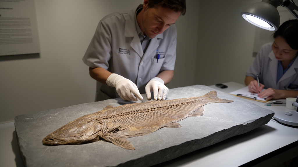
Using synchrotron X-ray microtomography, scientists have peered inside the fossilised pectoral fins of the Devonian stem lungfish Glyptolepis groenlandica and found something unexpected: a skeleton built by two different bone-forming processes at once, a combination the authors describe as remarkable.
Lungfishes first appeared in the Early Devonian, around 415 million years ago, and are regarded as the sister group to tetrapods, making them central to any reconstruction of how land vertebrates’ limbs evolved. Their early relatives rapidly diversified during the Devonian with well-ossified skeletons, but living lungfishes, represented by just three genera in Africa, South America and Australia, have postcranial skeletons that remain largely cartilaginous. Their fin skeleton has lost ossification altogether in modern forms. Understanding when and how that mineralisation was reduced has long depended on fossils such as Glyptolepis, whose ossified archipterygial fins, with a main bony axis running through the centre, preserve the microscopic traces of ancient development.
The fossils, collected from a Middle Devonian Givetian locality in Canning Land, East Greenland, each carry skulls between 8 and 9 centimetres long. Virtual thin sections and three-dimensional models were then generated with segmentation software, and a directional coloured lighting protocol revealed the orientation of internal bony struts, with longitudinal trabeculae appearing purple and transverse ones green.
The virtual histology of the first two specimens, NHMD 141677 and NHMD 152554, revealed a striking internal architecture. Each proximal mesomere, the segmental element of the archipterygial fin, carries an extremely thin outer cortex of bone, averaging just 120 micrometres thick in the diaphysis of the first specimen and only 85 micrometres in the second, thickening modestly in the ventral processes. Beneath this cortex lies an extensive spongy network of trabeculae surrounding a large central cavity filled with sediment during fossilisation. That cavity, the researchers concluded, was once occupied by cartilage that never mineralised and never fossilised. The evidence is subtle but compelling: three-dimensionally preserved vascular canals, roughly 100 micrometres in diameter, penetrate a few hundred micrometres into the sediment core and stop, never reaching the centre. If the core had been an empty medullary cavity, those canals would be floating with almost no connection to surrounding bone, and the flattened bodies of the fossils suggest hollow mesomeres would have collapsed during burial.
The trabeculae themselves tell a developmental story. In the metaphyses, the regions beneath the articular ends of each mesomere, the bony struts are longitudinally and conically oriented, while in the diaphysis, the shaft, they lose orientation and become more rounded. A third specimen, NHMD 153826, preserves mesomeres from the middle of the fin, and these show a reduced version of the same pattern: a cortex of merely 20 micrometres with no erosion bays, trabeculae concentrated only in the metaphyseal regions, and an almost trabecula-free diaphyseal centre. The authors interpret these middle mesomeres as an arrested stage of the same ossification process seen in the proximal elements, suggesting a developmental program in which mesomeres ossify to different degrees depending on their position along the fin.
Putting the specimens together, the team reconstructed a hypothetical sequence of mesomere development. A cartilaginous core first forms, surrounded by a perichondrium. Once the core nearly reaches its final size, a thin layer of perichondral bone is deposited around the diaphysis and over the dorsal and ventral processes. Blood vessels then invade the cartilage through erosion, forming canals at the core’s surface. In the diaphysis, erosion intensifies into bays beneath the inner cortical surface, where endochondral ossification generates a trabecular network. Crucially, unlike in most tetrapods, this erosion and bone formation remain confined to a layer no thicker than 400 micrometres beneath the cortex, leaving the central cartilage core intact. The longitudinal metaphyseal trabeculae, presumably formed by mineralisation around columns of aligned chondrocytes, indicate that the mesomere elongated during fin development, though their short length suggests that elongation was somewhat restricted.
The combination of ossification mechanisms is what makes the finding remarkable. In the diaphysis, the trabeculae appear to form through connective vascular sprouts developing between the perichondrium and the cartilage, inducing erosion and calcification of peripheral cartilage, a process otherwise seen only in the long bones of modern frogs and in the pectoral and pelvic girdles of extant coelacanths. In the metaphyses, however, endochondral ossification replaces the cartilaginous matrix surrounding columns of hypertrophic chondrocytes, the mechanism familiar from amniotes and some stem tetrapods. Notably, no primary centre of ossification, the trigger of classic long-bone development in tetrapods, ever formed in the cartilaginous core. The researchers propose that vascular invasion into the centre was inhibited, possibly because chondrocytes did not hypertrophy or the extracellular matrix was not degraded, keeping signalling factors such as vascular endothelial growth factor and Indian hedgehog from initiating ossification in the core.
To test how typical this pattern was among early sarcopterygians, the team compared Glyptolepis with coelacanths, a more phylogenetically basal lobe-finned lineage. The Triassic coelacanth Laugia groenlandica, unusual among early coelacanths in having well-ossified paired fins, shows an extensive trabecular network in its first pectoral mesomere, but one that has fully invaded the central core with no preferential orientation. That absence of oriented metaphyseal trabeculae implies the framework for long-bone-like elongation had not yet been established before the lungfish lineage split off, meaning mesomere elongation was probably present in the common ancestor of Dipnomorpha, the lungfish total group, and Tetrapodomorpha, but not at the sarcopterygian crown-group node. The living coelacanth Latimeria chalumnae, by contrast, has a fully cartilaginous pectoral fin whose cartilage lacks hypertrophic cells, confirming the group’s inability to elongate its fin skeleton, though canals similar to those in Glyptolepis pierce its girdle cartilage.
Because porolepiforms sit securely as the most rootward stem lungfishes, the pattern in Glyptolepis, a cartilage core ringed by a trabecular mesh, likely illustrates the primitive condition for the lungfish lineage. The authors note that the fossilised appendicular skeleton of the related genus Chirodipterus also shows an empty cavity at the centre of its mesomeres, suggesting that absence of primary ossification centres may have been widespread in early stem lungfish. In the extant lungfishes Protopterus annectens and Neoceratodus forsteri, cartilage cells in the centre of the mesomere core are distributed circularly while metaphyseal cells align longitudinally, a arrangement the researchers interpret as a lingering vestige of the ossification-inducing chondrocyte architecture of stem lungfishes like Glyptolepis.
The study, which is open access and based entirely on non-destructive imaging of museum specimens alongside comparative scans of larval and juvenile lungfish, coelacanths and a temnospondyl amphibian, positions Glyptolepis groenlandica as a reference point for tracing the evolutionary steps that stripped mineralisation from the modern lungfish skeleton. It also demonstrates that the elongation mechanism once considered a hallmark of tetrapod limb bones was already operating, in modified form, in the fins of stem lungfish. The team plans to extend this histological approach to other species of stem lungfish and coelacanths to refine the picture of how endochondral ossification evolved across the lobe-finned vertebrates that eventually walked onto land.
Subject of Research: Fossil histology of pectoral fin ossification in the Devonian stem lungfish Glyptolepis groenlandica
Article Title: Ossification of the pectoral fin of Glyptolepis groenlandica: implications for the evolution of the lungfish appendicular skeleton
Article References: Branigan, M. K., Bijl, S., Ahlberg, P. E., Tafforeau, P., Mansuit, R., Leyhr, J., Clement, A. M., Stundl, J., Dutel, H., Hirasawa, T., Haitina, T., & Sanchez, S. (2026). Ossification of the pectoral fin of Glyptolepis groenlandica: implications for the evolution of the lungfish appendicular skeleton. Developmental Biology Advances, 17(1), Article 16. https://doi.org/10.1186/s13227-026-00264-y
Image Credits: AI Generated
DOI: 10.1186/s13227-026-00264-y
Keywords: lungfish, Glyptolepis groenlandica, Devonian, endochondral ossification, pectoral fin, mesomeres, porolepiforms, synchrotron microtomography, paleohistology, evolution, sarcopterygians, Ossification
News Source: Drew Townsend. (October 30, 2026). Ancient Lungfish Fossil Reveals a Strange Hybrid of Bone-Forming Strategies. Scienmag.



