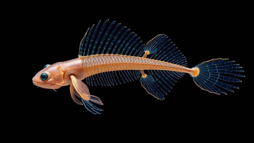
Paired appendages are one of the defining innovations of vertebrate life. The pectoral and pelvic fins of fishes and the forelimbs and hindlimbs of tetrapods transformed how vertebrates moved, fed, and colonized new environments, and they ultimately supplied the developmental scaffolding from which limbs evolved. Yet despite more than a century of comparative anatomy and decades of developmental genetics, a central question has remained stubbornly unresolved: how was the genetic system that builds paired appendages assembled in the first place?
Hox genes occupy a special place in developmental biology because their order along the chromosome mirrors the order of their expression along the head-to-tail axis of the embryo, a phenomenon known as colinearity. Genes at the posterior ends of the HoxA and HoxD clusters, including paralog groups 9 through 13, are expressed in the posterior trunk and tail regions of developing vertebrates, where they help assign identity to vertebrae, the hindgut, and the urogenital system. The new findings from zebrafish and the Iberian ribbed newt add a striking entry to this list: the same posterior Hox machinery that stamps identity onto the trunk also appears to be the trigger that first designates which cells will build the hindmost pair of appendages. This places the origin of hind appendages not at the level of morphological outgrowth, but at the level of cell fate specification, a stage that has been difficult to probe experimentally because the relevant cell populations are small, transient, and not yet visually distinct in the embryo.
The technical approach taken by the researchers reflects how modern developmental genetics has evolved to address such early events. In zebrafish, single mutations in Hox genes often produce subtle or no visible effects because of paralogy, the duplication of entire Hox clusters that occurred early in vertebrate history. Zebrafish carry duplicated Hox clusters inherited from a whole-genome duplication in the teleost lineage, meaning that a gene like hoxa9 exists in two copies, hoxa9a and hoxa9b, each capable of partially compensating for the loss of the other. Only by combining mutations across multiple paralogs — in this case five genes spanning both the HoxA and HoxD clusters — could the team reveal the full requirement for paralog group 9 and 10 function in pelvic fin formation. This combinatorial strategy mirrors what has been done in mice, where knocking out individual Hox genes rarely exposes their deepest roles, but cumulative disruption of paralog groups reveals dramatic transformations of body structure.
The complete loss of pelvic fins in zebrafish lacking these five genes is a particularly clean result because pelvic fins are the hindmost paired appendages in fish, directly analogous to hindlimbs in tetrapods. The fluorescent reporter lines used in the study allowed the researchers to visualize pelvic fin progenitor cells before any fin bud became visible under the microscope. When the five Hox genes were disrupted, these bilateral progenitor populations simply never appeared, indicating that the failure occurs at or before the stage when mesenchymal cells are first allocated to the pelvic appendage fate. This distinguishes the result from earlier work showing Hox requirements during fin or limb outgrowth and patterning, stages at which the appendage bud already exists and is growing. Here, the genes appear to act upstream of bud formation itself, at the point where a population of cells in the lateral plate mesoderm and adjacent tissue is being set aside to become the hind appendage.
The choice of the Iberian ribbed newt as a second experimental system is significant for reasons that go beyond simple taxonomic breadth. Newts belong to the salamander lineage, which is part of the lobe-finned vertebrate branch that also includes the ancestors of all tetrapods. Ray-finned fishes such as zebrafish and lobe-finned vertebrates such as newts diverged from a common ancestor more than 400 million years ago, making any shared developmental mechanism between them likely to have been present in that common ancestor. The newt experiments used CRISPR-Cas9 genome editing to disrupt all seven Hox9 and Hox10 paralogs simultaneously, a technically demanding feat in an organism with a long generation time and a large genome. The observation that animals losing hindlimbs also lost or markedly reduced expression of Prdm16, a transcription factor associated with hindlimb-forming regions, provides a molecular readout that connects the genetic disruption to a specific failure in progenitor specification rather than a general developmental defect.
Prdm16 itself is worth noting in this context. This gene encodes a chromatin-associated transcriptional regulator that has been studied primarily for its roles in adipocyte differentiation and in the development of the heart and facial structures. Its expression in prospective hindlimb-forming regions of the newt embryo, and its dependence on Hox9-10 function, suggests that it may act as a downstream effector or marker of the Hox-dependent program that establishes hind appendage progenitors. Whether Prdm16 is directly bound and regulated by Hox9-10 proteins, or whether it sits further downstream in a cascade, remains an open question that future experiments will need to address. What the current data establish is that the loss of Hox9-10 function produces a correlated loss of both the morphological appendage and a molecular marker of the appendage-forming field, strengthening the interpretation that the primary defect is in progenitor establishment.
The stepwise evolutionary model that the authors propose builds on a long history of comparative Hox biology. In insects and other protostomes, Hox genes pattern segment identity along the body axis but are not generally used to build paired appendages in the same way vertebrates deploy them. In vertebrates, the recruitment of posterior Hox genes into the appendage-patterning system has been inferred from several lines of evidence: the conserved expression of HoxA and HoxD cluster genes in developing fins and limbs across diverse species, the phenotypic consequences of Hox disruption in mice and fish, and comparative genomics showing conserved regulatory landscapes around the HoxD cluster that are active in appendages. What has been missing is direct functional evidence that the earliest step — the specification of the progenitor cells themselves — depends on posterior Hox genes, and that this dependence is shared across deeply diverged vertebrate lineages. The current study fills that gap.
The model suggests a sequence of events in the evolution of the vertebrate paired appendage system. First, an ancestral Hox9-10 program, perhaps originally functioning in trunk and tail patterning, was co-opted to designate cells for hind appendage formation. This step would have been sufficient to establish a basic posterior paired appendage. Second, additional posterior Hox genes from paralog groups 11 through 13 were incorporated into the program, expanding the patterning capacity to include proximal-distal and anterior-posterior axes within the growing appendage. Third, the expanded system was redeployed in anterior paired appendages, the pectoral fins and forelimbs, where Hox genes from paralog groups 1 through 8 also contribute to patterning in a reversed collinear fashion. This redeployment would have allowed the same core genetic machinery to build both pairs of appendages, a hallmark of the vertebrate body plan.
Supporting this model, previous comparative studies have shown that the HoxD cluster in vertebrates is flanked by regulatory elements that drive expression in developing appendages, and that these elements show signs of evolutionary conservation and modification across fish and tetrapod lineages. The HoxA cluster carries analogous regulatory landscapes. The integration of these regulatory architectures with the developmental functions now attributed to Hox9-10 in hind appendage progenitor specification provides a framework in which genetic innovation at the level of gene regulation could have progressively built the paired appendage system from simpler ancestral components.
It is important to note what the study does not claim. The authors are careful to state that their work does not directly reconstruct the evolutionary sequence of events that led to the modern paired appendage system. Developmental evidence in extant species can reveal which mechanisms are shared and therefore likely to be ancestral, but it cannot by itself demonstrate the order in which those mechanisms were assembled over deep time. Fossil evidence, paleontological records of fin and limb morphology in extinct vertebrates, and broader comparative genomic analyses will all be needed to complement the developmental data and test the stepwise model more rigorously. Nevertheless, the demonstration that Hox9-10 genes are required for hind appendage progenitor establishment in both a ray-finned fish and a lobe-finned salamander provides a strong indication that this function predates the divergence of these lineages and was likely present in the last common ancestor of jawed vertebrates.
The broader significance of this work extends into several areas of ongoing research. For developmental biologists, the identification of an early Hox-dependent step in hind appendage formation raises questions about the upstream signals that activate Hox9-10 in the relevant progenitor fields, and about the downstream targets through which Hox proteins execute their effects. For evolutionary biologists, the results provide a concrete molecular handle on the problem of how novel morphological structures arise through the modification of existing developmental programs rather than through the invention of entirely new genes. For researchers interested in regenerative biology, understanding how progenitor cells are specified for appendage formation in organisms with natural regenerative capacities, such as newts, could inform strategies for inducing regenerative responses in species that lack them.
Associate Professor Kawamura’s comments about the timeline for translating these findings into practical applications reflect a realistic assessment of where this research sits on the spectrum from basic discovery to applied science. The immediate value of the work is conceptual: it establishes a developmental connection between the posterior Hox code of the body axis and the Hox system used to build paired appendages, and it provides functional evidence for an ancestral posterior Hox program that contributed to the evolutionary assembly of the appendage-patterning system. Over the coming years, as genome editing tools become more refined and comparative developmental biology expands to include more diverse species, the principles uncovered here are likely to find relevance in studies of congenital limb malformations, in efforts to understand the genetic basis of appendage diversity across vertebrates, and in the broader quest to understand how the vertebrate body plan was assembled through evolutionary time.
The collaborative structure of the research team, drawing on expertise from Saitama University, Tottori University, the National Institute of Genetics, the National Institute for Basic Biology, and the Exploratory Research Center on Life and Living Systems, reflects the interdisciplinary nature of the questions being addressed. Genetic analysis in zebrafish requires facilities and expertise in fish husbandry, transgenesis, and imaging. CRISPR-Cas9 disruption in newts requires specialized knowledge of salamander embryology and genome editing in an organism with a genome roughly ten times the size of the human genome. Skeletal imaging, fluorescent reporter design, and comparative analysis across species each demand their own technical competencies. The integration of these approaches within a single study demonstrates how modern research into fundamental questions of body plan evolution increasingly depends on teams that span multiple institutions and multiple model systems.
As the field moves forward, several experimental directions follow naturally from this work. Identifying the direct transcriptional targets of Hox9-10 proteins in the pelvic fin progenitor field of zebrafish would help define the gene regulatory network downstream of the Hox code. Testing whether the same Hox9-10 requirement applies to pectoral fin or forelimb progenitors, or whether anterior appendages rely on a different set of Hox genes at the progenitor stage, would clarify the degree to which the ancestral program was indeed posterior-specific before being redeployed anteriorly. Extending the analysis to additional vertebrate lineages, including cartilaginous fishes and more basal ray-finned fishes, would help pinpoint when in vertebrate evolution the Hox9-10-dependent hind appendage program was established. Each of these directions builds on the foundation laid by the current study and contributes to the larger goal of understanding how one of the most consequential innovations in vertebrate history came to be.
Subject of Research: Evolutionary developmental biology of Hox genes and vertebrate paired appendages
Article Title: Ancient Hox code reveals the developmental roots of vertebrate hind fins and limbs
Article References: Ancient Hox code reveals the developmental roots of vertebrate hind fins and limbs. Original publication (Bibliographic details unavailable.)
Image Credits: AI Generated
DOI: Not provided
Keywords: Hox genes, zebrafish, Iberian ribbed newt, pelvic fins, hindlimbs, evolutionary developmental biology, CRISPR-Cas9, PNAS, Ancient, code, reveals, developmental
News Source: Juliet Wilcox. (October 19, 2026). Ancient Hox9–10 genes reveal the hidden origins of vertebrate hind fins and limbs. Scienmag.