For decades, developmental biologists have recognised only two places in the vertebrate head where ectoderm and endoderm meet directly, without intervening mesoderm: the primary mouth and the pharyngeal gill slits. A new study, published as an open-access research article in Developmental Biology Advances on 4 October 2026, documents a third such interface and describes in cellular detail how it forms on the surface of the embryonic face in bichir, sturgeon and gar embryos.
The interface involves the pre-oral gut, or POG, an endodermal structure that appears in early embryos of bichir, sturgeon and gar. The authors had previously identified this ecto-endodermal contact in these species, showing that its developmental origin precedes both the formation of the primary mouth and the opening of the pharyngeal slits. In the new work, they set out to characterise the ecto-endodermal dynamics during POG externalisation, a process that had remained largely unknown at the cellular level.
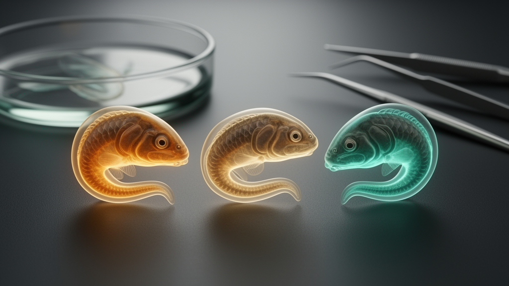
Using lineage tracing, high-resolution microscopy and immunohistochemistry, the team followed the interactions between the two germ layers as the POG was pushed onto the embryonic surface in bichir, gar and sturgeon embryos. The results reveal that the three species achieve this externalisation in strikingly different ways, reflecting different fates and functions of the structure in each lineage.
In bichir and gar, the process is comparatively simple: the endodermal layer bulges outward, and the exposed endodermal secretory cells come to sit on the embryonic surface, where they constitute the animals’ cement glands. These glands serve an adhesive function, anchoring the embryo. In sturgeon, by contrast, the study revealed an intricate intermixing between the POG and the surface periderm cells, with periderm cells alternating with secretory endodermal cells and expanding the hatching gland cells over the ventral rostrum.
The functional outcome differs accordingly. In sturgeon, the POG gives rise to transient hatching gland cells that accumulate and then release hatching enzymes. In bichir and gar, the POG instead forms cement glands with adhesive function. The authors report that these changes in the cellular structure of the POG reflect its different fate and function in the species studied, and they document substantial changes in the composition of the endodermal secretory cells across development.
The work also carries implications for how the germ layer origin of these rostral, ephemeral embryonic glands is understood across vertebrates. The authors compare the various architectures of the ectoderm-endoderm interfaces in the three species and discuss possible changes in the germ layer origin of these transient glands, an indication that what appears to be a conserved embryonic structure may have shifted its cellular makeup over evolutionary time.
Because the interface forms within the pre-mandibular head domain, ahead of the other oropharyngeal openings, it represents a uniquely early ecto-endodermal contact in the developing head. The study’s conclusions emphasise the complex dynamics and architecture of this interface, which had been overlooked in the classical two-site model of direct ecto-endodermal interaction in the vertebrate head.
It is worth noting the scope of the findings. The observations come from embryos of three non-teleost fish species — bichir, sturgeon and gar — examined with lineage tracing, microscopy and immunohistochemistry. The article is being shared early to provide faster access to peer-reviewed, accepted research; it is citable and carries a permanent DOI, but this version is subject to further edits and will be replaced by the final Version of Record. The authors include A. Huysseune, A. Horackova, T. Suchanek, M. Minařík, V. Klukanová, R. Franěk, L. Arias-Rodriguez and R. Cerny, with the article reporting 79 accesses and an Altmetric score of 1 at the time of the source record.
For developmental biologists, the study adds a well-characterised example of how ectoderm and endoderm can interact directly in the head without mesoderm between them, and it shows that the architecture of such interfaces can differ substantially among the species studied.
Subject of Research: Ectoderm-endoderm interactions during externalisation of the pre-oral gut in bichir, gar and sturgeon embryos
Article Title: The pre-oral gut on the embryonic face reveals novel ectoderm-endoderm interactions during vertebrate head development
Article References: Huysseune, A., Horackova, A., Suchanek, T., Minařík, M., Klukanová, V., Franěk, R., Arias-Rodriguez, L., & Cerny, R. (2026). The pre-oral gut on the embryonic face reveals novel ectoderm-endoderm interactions during vertebrate head development. Developmental Biology Advances. https://doi.org/10.1186/s13227-026-00272-y
Image Credits: AI Generated
DOI: 10.1186/s13227-026-00272-y
Keywords: pre-oral gut, ectoderm-endoderm interface, vertebrate head development, bichir, sturgeon, gar, cement glands, hatching gland, periderm, embryonic development, lineage tracing, endoderm
News Source: Drew Townsend. (October 28, 2026). An Unexpected Ectoderm-Endoderm Interface Emerges on the Embryonic Face of Bichir, Gar and Sturgeon. Scienmag.