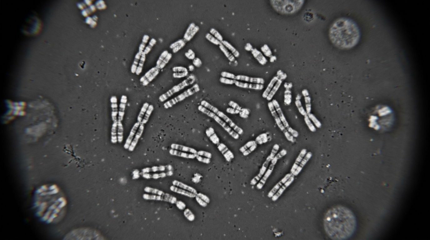
Genome engineers have long been able to cut, delete and rewrite stretches of DNA with remarkable precision, but one of the most common forms of genomic change has remained stubbornly out of reach: duplication. Now, researchers report a protocol for amplification editing, a genome-editing strategy that enables efficient and precise duplication of genomic sequences ranging from tens of base pairs to chromosomal scales, and they lay out detailed procedures for designing, executing and analyzing the technique in mammalian cells. The work, published in Nature Protocols, addresses a gap that has persisted even as CRISPR-based tools have transformed the manipulation of genomes over the past decade.
Duplication events are fundamental to evolution and contribute to complex genomic disorders. Repetitive sequences make up more than half of the human genome, and among these, telomeres and centromeres are essential for maintaining genome stability, while some other repeats are involved in the development of human diseases. Gene dosage regulation depends heavily on how many copies of a gene a cell carries, and duplications contribute to the development of various human diseases including cancer and inherited disorders. Despite the critical roles of gene duplication and repeated sequences, the tools available to engineer them deliberately have lagged far behind those for other types of edits.
Traditional Cas9 nucleases, when combined with pairs of single-guide RNAs, can generate large-scale genomic rearrangements such as deletions, inversions and inter-allelic recombination or chromosomal translocations by targeting distinct sites. However, this approach often results in complex and heterogeneous outcomes with low efficiency and limited precision, in which duplications appear only as rare by-products. Next-generation tools improved matters considerably. Base editing couples a deaminase to a catalytically impaired Cas protein to introduce precise point mutations such as C-to-T or A-to-G. Prime editors integrate a Cas9 nickase fused to a reverse transcriptase with a prime editing guide RNA that carries a primer binding site and a reverse transcriptase template, enabling precise installation of base substitutions, small deletions and insertions without generating double-strand breaks or requiring donor DNA templates. Paired pegRNAs even allow precise generation of large-scale genomic deletions, insertions and inversions. Yet methods for engineering genomic structural variations, especially DNA duplication, remained lacking.
Amplification editing, abbreviated AE, fills that void with an elegant molecular trick. The system uses a Cas9 nickase fused to a reverse transcriptase, known as prime editor 2, together with a pair of specially designed pegRNAs. These pegRNAs target opposite DNA strands in a protospacer-adjacent motif, or PAM, out orientation, with the PAMs positioned outside the intended duplication region. Each pegRNA directs the nickase to introduce a nick at the region of interest, generating single-strand breaks precisely at both ends of the targeted sequence. The reverse transcriptase templates are designed to be fully or partially complementary, and two short complementary DNA 3-prime flaps are generated during reverse transcription.
Those flaps are the heart of the mechanism. The two flaps can anneal through base pairing to form a short duplex, which exposes a free 3-prime hydroxyl group that functions as a primer for DNA synthesis. The endogenous DNA polymerase or the reverse transcriptase of the prime editor is then recruited to extend from the 3-prime end of the duplex. During this process, the polymerase unwinds the genomic DNA and uses one strand of the intervening sequence as the template. DNA synthesis proceeds across the region between the two nick sites and duplicates the intervening genomic sequence. After DNA synthesis is completed, cellular DNA repair pathways seal the two nicks. As a result, the sequence between the two nick sites is duplicated, with a short sequence derived from the RTT flaps retained at the junction between the duplicated region. For megabase-level sequence duplication, the authors speculate that the endogenous DNA replication machinery, including the replication fork, is involved.
The design choices matter enormously to performance. Spacer sequences, selected with tools such as CRISPOR, are a major determinant of AE efficiency because nick generation depends primarily on them. When duplicating a 1-kilobase fragment at different loci, editing efficiency can differ by as much as 1.68-fold, with the RUNX1 locus reaching 47.77 percent compared with 28.47 percent at AAVS1. Shorter distances between the two PAM sites generally contribute to higher editing efficiency, though this parameter is constrained by the size of the desired duplicated region and the availability of suitable PAM sequences. Flap length has a stronger effect than GC content: 10-base-pair flaps show little or no detectable editing, while 30-to-50-base-pair flaps generally exhibit higher efficiencies, and flap sequences containing 30 to 80 percent GC are compatible across the tested loci. Unless a specific junction sequence is required, the researchers recommend a previously validated 30-base-pair sequence, TCAGTAACCCGGAGCTCTAATTCTCCCGAA, which performed robustly across multiple genomic loci and cell lines. Because AE requires a pair of pegRNAs, the protocol advises designing three to five pegRNAs for each region and screening all possible combinations; in the team’s previous study, AE products were detected in 27 of 32 randomly selected pegRNA-pair combinations across eight loci, although efficiencies varied substantially.
Quantifying the results requires a layered toolkit. Junction detection relies on in-in PCR, in which two primers positioned within the duplicated region span the newly formed junction, followed by agarose gel electrophoresis and Sanger sequencing. Editing efficiency is measured independently by droplet digital PCR, which divides each reaction into tens of thousands of individual droplets and achieves absolute quantification sensitive enough to detect rare editing events at frequencies below 1 percent, and by unique molecular identifier-based next-generation sequencing, which labels each DNA molecule with a 12-nucleotide barcode to eliminate PCR amplification bias. Junction purity is assessed through a two-round PCR with barcodes and Illumina adapters, measuring the fraction of reads that exactly match the desired junction. For validation, out-out PCR and Sanger sequencing confirm small duplications under 1 kilobase, Oxford Nanopore long-read sequencing verifies duplications up to 10 kilobases, and duplications exceeding 100 kilobases are characterized by whole-genome sequencing and fluorescence in situ hybridization, in which additional probe signals and chromosome elongation visually confirm large-scale events.
The reported performance figures are striking. At the HEK3-SC locus, a 234-base-pair duplication was generated at an efficiency of 69.73 percent as measured by ddPCR, with a comparable 79.45 percent obtained by UMI-based NGS. In bulk edited cells, small fragments are prone to undergo multiple duplications: at the HEK4 locus in HEK293T cells, duplication of a 262-base-pair fragment increased the copy number of the targeted region from one to a range of 2 to 14, with an average of 4.2 copies, while duplication of a 7,677-base-pair fragment at RUNX1 increased the copy number from one to two. This variation is stochastic and heterogeneous, associated with editing efficiency and duplicated-region length, but it does not prevent isolation of single-cell clones with defined copy numbers through downstream screening. In validated single-cell clones, the researchers have not observed copy-number loss or rearrangement during routine expansion across different cell lines, though they recommend periodic re-characterization during prolonged culture because instability under stressful conditions has not yet been evaluated.
The delivery and scope of the system are unusually broad. AE has been performed with high efficiency across diverse cell types, including human cell lines such as HEK293T, Huh-7, U2OS and K562, diploid human cell lines including RPE-1 and diploid HAP1, human embryonic stem cells, and primary human T cells, as well as mouse cell lines including N2a, haploid and diploid mouse embryonic stem cells. Plasmid delivery by electroporation or lipid transfection is specified for each cell type, and for primary T cells, STING-targeting siRNAs should be electroporated three days before AE delivery to mitigate cytotoxicity triggered by electroporation-mediated DNA delivery. Most AE experiments can be completed within 2 weeks, and the full workflow from pegRNA construction through validation of megabase-scale products spans roughly three to four weeks for the integrity-checking stage.
Compared with tandem duplication prime editing, a recently developed method that uses paired pegRNAs to generate 3-prime flaps homologous to genomic sequences and mediates tandem duplications up to 10 kilobases, AE differs by introducing a pair of complementary flaps rather than homology with genomic sequences, and it generates duplications beyond 10 kilobases, achieving precise duplications up to the 100-megabase level. Alternative PAM-out designs using pegRNA-sgRNA or sgRNA-sgRNA pairs produced duplications only at low efficiency and were less flexible in design. The limitations are candidly acknowledged: small-fragment duplications are feasible in non-dividing cells, whereas large-fragment duplications may depend on endogenous DNA polymerases and require cell cycle progression, potentially limiting chromosomal-scale duplication in non-dividing cells, and a short junction scar may be retained when a randomly generated RTT sequence is used, which could be problematic in protein-coding regions or regulatory elements where scarless RTT design is preferred but may reduce design flexibility and compromise editing efficiency. The researchers have used the protocol to restore gene expression, enhance endogenous microRNA expression and increase HBA levels as well as alpha-over-gamma-globin ratios in a cellular model, and they point to applications in modeling copy-number variations in cancer and developmental disorders, generating disease models in established cell lines and primary cells, creating animal models of chromosomal microduplication diseases, and exploring potential for gene-based therapy. For researchers in genetics, synthetic biology, cancer biology, developmental biology and translational medicine, a programmable duplication tool of this reach opens doors that double-strand-break methods never could.
Subject of Research: A genome-editing protocol for duplicating genomic sequences in mammalian cells
Article Title: Efficient and precise large-scale genomic duplication using amplification editing
Article References: Xie, S.-S., He, Z., & Yin, H. (2026). Efficient and precise large-scale genomic duplication using amplification editing. Nature Protocols. https://doi.org/10.1038/s41596-026-01454-7
Image Credits: AI Generated
DOI: 10.1038/s41596-026-01454-7
Keywords: amplification editing, genome editing, prime editing, pegRNA, genomic duplication, structural variation, gene dosage, ddPCR, whole-genome sequencing, FISH, HEK293T, CRISPR
News Source: Juliet Wilcox. (November 5, 2026). Amplification Editing Duplicates Genomic Sequences from Base Pairs to Chromosomal Scale. Scienmag.