In the relentless pursuit to advance cancer diagnosis and treatment, researchers at the University of Auckland and its affiliated institutions have unveiled an illuminating review that charts the evolution of artificial intelligence (AI) in automating the segmentation of meningiomas from magnetic resonance imaging (MRI) scans. Meningiomas, tumors originating from the meninges—the delicate protective tissues enveloping the brain—pose diagnostic challenges due to their varied size and morphology. Manual identification and delineation on MRI images not only demand considerable time but also suffer from inter-observer variability, especially with minute or irregularly shaped tumors. The advent of AI, particularly deep learning methodologies, has ushered in a transformative era, enabling rapid, standardized, and precise tumor detection that holds profound implications for clinical decision-making and patient management.
This comprehensive review by scientists from the University of Auckland, Auckland City Hospital, and the Matai Medical Research Institute scrutinizes 34 pivotal studies published between 2020 and 2025. These studies collectively delve into AI-driven frameworks tasked with automatic meningioma segmentation using MRI data. The review meticulously dissects advancements in model architectures, evaluates the impact of dataset diversity and imaging modalities, and identifies bottlenecks hindering broader clinical deployment. Published in the peer-reviewed journal Brain Network Disorders, this synthesis not only highlights current triumphs but also calls for strategic innovation to propel AI applications from research settings into everyday medical practice worldwide.
Core to their findings is the pronounced influence of model architecture on segmentation performance, transcending traditional expectations that larger datasets and higher image quality unequivocally lead to better outcomes. The team emphasizes that while contrast-enhanced T1-weighted MRI emerges as the gold standard imaging protocol for meningioma visualization, enhancements in AI model design—incorporating sophisticated hybrid frameworks and intricate feature extraction techniques—have yielded the most significant accuracy improvements. This reframing challenges prevailing assumptions in radiological AI development, underscoring the nuanced interplay between data and model sophistication.
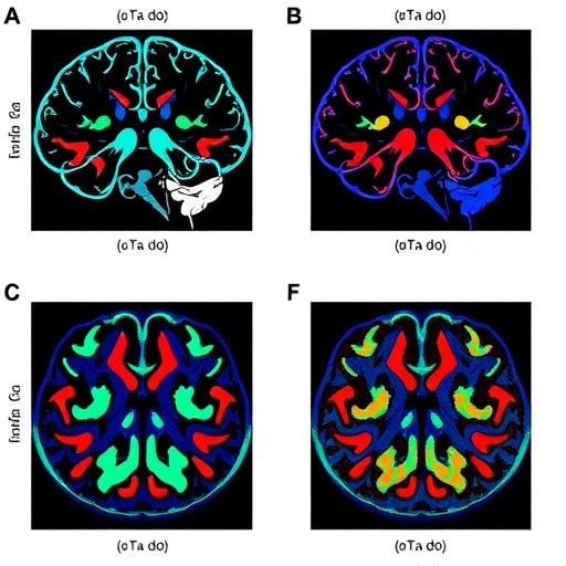
Quantitative evaluation of segmentation proficiency primarily used the Dice similarity coefficient, an established metric assessing the degree of overlap between predicted tumor boundaries and expert annotations. The reviewed AI models stratified into three distinct tiers based on Dice scores. Tier 1, exemplified by architectures like DeepLabV3+, attained near-perfect overlaps with scores nearing 0.98, leveraging extensive datasets such as the 3,064-image collection from Figshare. However, their computational intensity presents practical constraints. Tier 2 models, including variants of the widely adopted 2D U-Net, offer a compelling balance with Dice scores ranging from 0.8 to 0.9, making them attractive for routine clinical use. Conversely, Tier 3 models prioritize operational speed and efficiency at the expense of some accuracy, fitting scenarios requiring rapid assessments with moderate precision.
Dr. Hamid Abbasi, the lead investigator and a senior research fellow at the Auckland Bioengineering Institute and Center for Brain Research, elucidates the unexpected trajectory of AI advancements: “Our analysis revealed that smarter AI model architectures have propelled segmentation quality more decisively than the sheer volume of training data or enhancements in MRI scan fidelity. This insight recalibrates how we approach AI design for complex medical imaging tasks.” This paradigm shift facilitates targeted innovation focusing on algorithmic refinement rather than merely amassing data repositories.
Remarkably, state-of-the-art AI systems demonstrate remarkable efficiency, processing complex segmentation tasks in timescales as swift as 15 seconds—orders of magnitude faster than manual delineations conducted by radiologists. Nima Sadeghzadeh, the first author of the study, expresses optimism about this leap, emphasizing the promise AI holds in augmenting clinical workflows by delivering highly accurate, consistent tumor maps with unprecedented speed, thereby expediting diagnosis and enabling timely treatment interventions.
Despite these advances, challenges persist. Small-volume tumors below 3 milliliters frequently evade detection, raising concerns about missed diagnoses in early-stage meningiomas. Moreover, the substantial computational demands of tier 1 models pose integration hurdles in resource-limited hospitals where high-performance computing infrastructure remains scarce. Most existing AI models also grapple with generalizability, displaying diminished performance when applied to external datasets from institutions with differing imaging protocols or patient demographics, thereby restricting universal clinical adoption.
Addressing these barriers calls for concerted research efforts aimed at developing universally robust AI models capable of seamless adaptation across heterogeneous medical environments. Emphasis on model efficiency—enabling operation on modest hardware without compromising accuracy—and enhancing dataset diversity to encompass varied clinical scenarios are pivotal steps. Such progress would democratize access to AI-powered diagnostic tools, bridging disparities between advanced medical centers and under-resourced facilities globally.
The synthesis further advocates for rigorous benchmarking standards and collaborative data-sharing initiatives to catalyze transparency and reproducibility in AI research. Establishing consensus on best practices for MRI acquisition, annotation protocols, and evaluation metrics can harmonize efforts, accelerating innovation and regulatory acceptance. This holistic framework is indispensable in transforming the promise of AI from experimental modalities to validated medical devices entrusted with patient care.
Fundamentally, this review underscores the symbiotic relationship between computational innovation and clinical acumen in confronting complex neurological disorders. By harnessing AI’s capability to decode intricate imaging patterns otherwise imperceptible to the human eye, clinicians can achieve earlier, more reliable meningioma detections that profoundly influence prognostic trajectories. As these technologies evolve, their integration within multidisciplinary treatment paradigms heralds a new epoch in personalized neuro-oncology.
In conclusion, the journey from manual brain tumor segmentation to automated AI-driven delineation epitomizes the transformative potential of artificial intelligence in medicine. The University of Auckland-led review not only chronicles the state-of-the-art progress but also charts a clear roadmap for future research priorities. Overcoming extant limitations will unlock widespread clinical adoption, ultimately improving outcomes for patients worldwide through faster diagnosis, refined treatment planning, and enhanced monitoring of meningiomas.
Subject of Research:
Artificial intelligence applications in medical imaging, specifically automated segmentation of meningioma brain tumors from MRI scans.
Article Title:
Artificial intelligence-driven advances in automatic segmentation of meningioma brain tumors: A systematic review
News Publication Date:
March 24, 2026
Web References:
https://doi.org/10.1016/j.bnd.2026.01.001
References:
Abbasi, H., Sadeghzadeh, N., et al. (2026). Artificial intelligence-driven advances in automatic segmentation of meningioma brain tumors: A systematic review. Brain Network Disorders, 2(1). https://doi.org/10.1016/j.bnd.2026.01.001
Image Credits:
Nima Sadeghzadeh and Hamid Abbasi, University of Auckland, New Zealand
Keywords:
Meningioma, Artificial Intelligence, Deep Learning, MRI, Brain Tumor Segmentation, Medical Imaging, Neural Networks, DeepLabV3+, U-Net, Dice Score, Radiology, Neuro-oncology
Tags: advances in brain tumor AI modelsAI brain cancer segmentationAI challenges in cancer imagingAI clinical decision supportAI in neuro-oncologyautomatic meningioma detectionbrain network disorder researchbrain tumor diagnosis AIdeep learning MRI analysismedical image segmentation AImeningioma tumor imagingMRI-based tumor delineation

