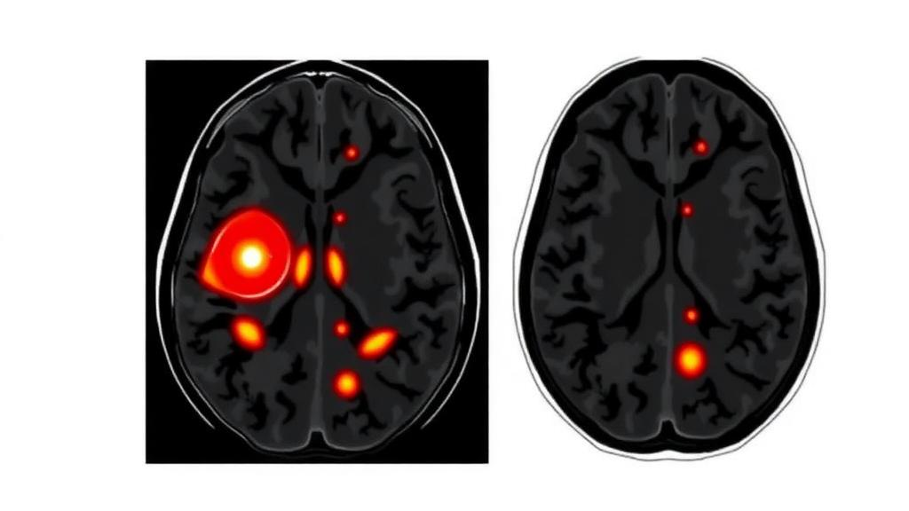
Prostate cancer is a disease of degrees. Two men can carry tumors in the same gland, yet one may live for decades without treatment while the other needs immediate, aggressive intervention. The difference lies in how disordered the cancer cells have become, a quality that pathologists capture in the Gleason score and its modern successor, the ISUP grade group. The problem is that determining that grade has almost always required a needle. Now, a team of radiologists and researchers in Nanjing, China, has shown that a machine learning model can predict these grade groups noninvasively, before any biopsy, by teaching an algorithm to see the hidden internal architecture of prostate tumors on routine MRI scans.
The study, published in BMC Medical Imaging, drew on 412 patients with pathologically confirmed prostate cancer treated at two medical centers between July 2021 and August 2024. The researchers divided the patients into a training cohort of 196 men, an internal validation cohort of 82, and a fully independent external validation cohort of 134 from a second hospital. This three-way split matters. Many artificial intelligence studies in medicine perform impressively on the data they were trained on and then collapse when confronted with patients from a different scanner, a different protocol, or a different population. By testing on an external cohort, the Nanjing team deliberately exposed their model to the harsher reality of clinical practice.
The technical heart of the work is something called habitat imaging. A tumor is not a uniform blob; it is a patchwork of regions with different cell densities, blood supplies, and degrees of cell death. Conventional radiomics treats the whole tumor as one object and averages away much of this internal diversity. Habitat analysis does the opposite. Using the K-means clustering algorithm, the researchers automatically partitioned each tumor into subregions, or habitats, that share similar signal characteristics on the two sequences that make up biparametric MRI: T2-weighted imaging, which reveals tissue structure, and diffusion-weighted imaging, which measures how freely water molecules move, a proxy for cellular crowding. Radiomic features, hundreds of quantitative descriptors of texture, shape, and intensity, were then extracted separately from each habitat subregion.
From this feature bank, the team built forty predictive models using five different machine learning algorithms, combining clinical variables, conventional imaging features, and habitat-derived radiomics in various configurations. The winner was a comprehensive model that integrated clinical factors, standard imaging features, and radiomic features drawn specifically from the third habitat subregion of the T2-weighted images. Its performance, measured by the area under the receiver operating characteristic curve, or AUC, reached 0.867 in the training cohort, 0.900 in internal validation, and 0.826 in the external validation cohort. An AUC of 1.0 represents perfect discrimination, 0.5 represents a coin flip, and values in the 0.8 to 0.9 range are generally considered good to excellent for a diagnostic model. The fact that performance held up, with only modest degradation, in the external cohort is the study’s most convincing credential.
Two clinical variables emerged as independent predictors in their own right. The first was total prostate-specific antigen, or tPSA, the familiar blood marker whose levels rise as prostate disease progresses; each unit increase carried an odds ratio of 1.041, a small but statistically robust effect. The second was more novel: the ADC ratio, calculated as the mean apparent diffusion coefficient of the tumor divided by that of normal prostate tissue. Because cancerous tissue is densely packed with cells, water diffuses less freely there, lowering the ADC value. A ratio far below one signals a tumor that is restricting water movement relative to its healthy surroundings, and in this study it was an extremely strong predictor of high-grade disease, with an odds ratio so small it rounded to less than 0.001.
What sets this study apart from much of the radiomics literature is its commitment to interpretability. Black-box models have justifiably made clinicians wary: if an algorithm says a tumor is aggressive but cannot explain why, few urologists will act on it. The researchers therefore applied SHAP analysis, short for SHapley Additive exPlanations, a technique borrowed from cooperative game theory that assigns each input feature a precise contribution to every individual prediction. With SHAP, the model’s reasoning can be visualized feature by feature, showing exactly which habitat characteristics and clinical values pushed a given patient’s prediction toward a high or low grade group. This transparency does not just build trust; it also gives researchers a way to check whether the model is learning genuine tumor biology or exploiting scanner artifacts.
Discrimination alone is not enough for a model to be clinically useful, and the team knew it. They therefore ran calibration curves, which test whether a model’s predicted probabilities match actual outcomes, and decision curve analysis, which quantifies the net benefit of acting on the model’s predictions across the full range of clinical decision thresholds. The comprehensive model outperformed all thirty-nine alternatives on both measures, achieving superior calibration and greater clinical net benefit. In practical terms, this means the model does not merely rank patients correctly; its probability estimates are trustworthy enough to inform real decisions about who needs immediate treatment and who might safely be monitored.
The clinical stakes are considerable. ISUP grade group determination currently depends on biopsy, an invasive procedure that samples only a fraction of the gland and can both undergrade and overgrade tumors. A reliable preoperative estimate from an MRI scan that patients already undergo could help urologists individualize their recommendations, distinguishing men who need radical treatment from candidates for active surveillance, and could inform surgical planning before tissue is ever obtained. The authors are careful to position the model as an auxiliary tool rather than a replacement for pathology, a framing that reflects both scientific honesty and the current regulatory climate for medical AI.
There are, of course, caveats. The study is retrospective, meaning the researchers worked backward from existing patient records rather than prospectively enrolling patients, and the requirement for informed consent was waived accordingly, with institutional review board approval and full anonymization. Both participating centers are in China, and the model was trained on biparametric protocols from a specific three-year window; generalization to other populations, scanners, and imaging protocols remains to be demonstrated. The funding came from the Innovation and Development Fund of Jiangsu Hospital of TCM, and the authors declare no competing interests. The article was published open access on October 6, 2026, as a citable early version subject to final editorial edits.
Even with those limitations, the work points toward a broader shift in medical imaging. Rather than asking radiologists to eyeball tumors and guess, habitat analysis decomposes each lesion into its constituent microenvironments and lets interpretable machine learning weigh the evidence. The combination of K-means habitat segmentation, SHAP-based transparency, and rigorous external validation offers a template that other research groups can follow for cancers far beyond the prostate. If future prospective studies confirm these results, the humble MRI scan could become something closer to a virtual biopsy, grading tumors from the inside out without a single needle.
Subject of Research: Noninvasive prediction of prostate cancer ISUP grade groups using MRI habitat radiomics and interpretable machine learning
Article Title: MRI habitat analysis using interpretable machine learning models for ISUP grade group prediction in prostate cancer
Article References: Chen, S., Wang, Y., Chen, H., Cui, J., Jiang, K., Shen, L., & Chen, J. (2026). MRI habitat analysis using interpretable machine learning models for ISUP grade group prediction in prostate cancer. BMC Medical Imaging. https://doi.org/10.1186/s12880-026-02870-7
Image Credits: AI Generated
DOI: 10.1186/s12880-026-02870-7
Keywords: prostate cancer, MRI, habitat imaging, radiomics, machine learning, ISUP grade group, biparametric MRI, SHAP, ADC ratio, K-means clustering, predictive medicine, BMC Medical Imaging
News Source: Nathaniel Bowman. (October 6, 2026). AI Reads Tumor Habitats on MRI to Predict Prostate Cancer Aggressiveness Before Surgery. Scienmag.