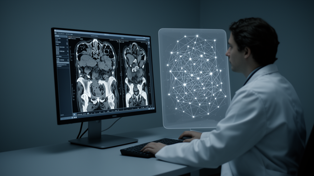
Bladder cancer is one of the most common malignancies of the urinary tract, and its treatment increasingly depends on molecular details that traditionally require a biopsy. Now, a team of researchers in China has shown that a sophisticated form of artificial intelligence image analysis, applied to routine computed tomography urography scans, can predict the HER2 status of bladder tumors before any tissue is taken. The study, published in BMC Medical Imaging, suggests that the information clinicians need to guide targeted therapy may already be hiding inside standard diagnostic images.
HER2, short for human epidermal growth factor receptor 2, is a protein that drives growth in some cancers and has become a critical therapeutic target. In bladder cancer, HER2 overexpression or amplification identifies patients who may respond to antibody-drug conjugates such as disitamab vedotin, a drug that has shown promise in HER2-positive urothelial tumors. The problem is that determining HER2 status currently requires immunohistochemistry or fluorescence in situ hybridization on tissue samples obtained through transurethral resection or biopsy. These procedures are invasive, carry procedural risks, sample only part of a heterogeneous tumor, and cannot always be repeated easily to track how the molecular profile changes over time.
The research team, led by investigators from Shaoxing University, Shaoxing People’s Hospital, and Zhuji People’s Hospital in Zhejiang Province, took a different approach. They turned to radiomics, a technique that extracts hundreds of quantitative features from medical images, features that are invisible to the human eye but encode information about tumor texture, shape, intensity distribution, and spatial patterns. When these features are fed into machine learning algorithms, they can reveal subtle relationships between image appearance and underlying biology, including molecular characteristics that pathologists normally detect only under the microscope.
The study enrolled 190 patients with pathologically confirmed bladder cancer who had undergone preoperative computed tomography urography, known as CTU, between October 2019 and September 2024. CTU is a multiphase CT scan of the urinary tract that captures images at different time points after contrast injection, including corticomedullary, nephrographic, and excretory phases, each highlighting different tissue characteristics. All patients had known HER2 status determined by standard pathological testing, providing the ground truth against which the imaging models could be validated.
To build the predictive models, the researchers first delineated regions of interest covering the tumors on each CTU phase and extracted a large panel of radiomic features, including those based on gray-level run length matrices, gray-level co-occurrence matrices, and gray-level size zone matrices. These mathematical descriptors quantify patterns such as coarseness, contrast, homogeneity, and directional texture within the tumor image. Features were filtered using the Least Absolute Shrinkage and Selection Operator, or LASSO, a regression method that shrinks less informative coefficients toward zero and effectively selects the most predictive variables while guarding against overfitting.
The cohort was randomly divided into a training set of 133 patients and an independent test set of 57 patients. Five different machine learning classifiers were then trained on the selected features: logistic regression, support vector machine, k-nearest neighbors, extreme gradient boosting, and random forest. Among these, the random forest model built on corticomedullary-phase images emerged as the clear winner, achieving an area under the receiver operating characteristic curve of 0.798 in the training set and 0.734 in the test set. The corticomedullary phase, which captures the early arterial enhancement of renal cortex and tumor vasculature, appears to contain the richest information about tumor biology relevant to HER2 expression.
The researchers did not stop at imaging features alone. They integrated the radiomic signature with clinical parameters to build a combined clinical-radiomics model, and the results were striking. This hybrid model achieved an AUC of 0.876 in the training cohort and 0.763 in the test cohort, outperforming the radiomics-only approach. Model performance was further evaluated using calibration curves, which assess how well predicted probabilities match observed outcomes, and decision curve analysis, which quantifies the clinical net benefit of using the model at various decision thresholds. Both analyses confirmed that the combined model offered meaningful clinical utility.
Among the 190 patients, 118, or 62.1 percent, were HER2-positive, reflecting the substantial proportion of bladder cancers that harbor this targetable alteration. Patient ages ranged from 26 to 90 years, with median ages of 72 in the HER2-positive group and 68 in the HER2-negative group. The relatively high prevalence of HER2 positivity in this cohort underscores why a non-invasive screening approach could have broad clinical impact, since a large fraction of patients might be eligible for HER2-directed therapies if their status could be reliably determined before treatment planning.
The implications of this work extend beyond bladder cancer. Radiomics-based prediction of molecular markers represents a growing frontier in precision oncology, sometimes described as radiogenomics or imaging genomics. If validated in larger, multicenter cohorts, this approach could allow clinicians to triage patients for HER2-targeted therapy using images already acquired for diagnosis, reducing the need for repeated invasive procedures and enabling faster treatment decisions. It could also be applied longitudinally, tracking molecular evolution of tumors over the course of therapy without additional biopsies.
The authors acknowledge that this was a single-center retrospective study, and external validation across diverse populations and scanner platforms will be essential before clinical adoption. Standardization of image acquisition and feature extraction, guided by initiatives such as the Image Biomarker Standardization Initiative, remains a key challenge for the field. Nevertheless, the demonstration that a routine CTU scan, combined with machine learning, can predict HER2 status with acceptable diagnostic performance marks a significant step toward non-invasive molecular profiling of bladder cancer, bringing the promise of personalized, image-guided oncology closer to everyday practice.
Subject of Research: Radiomics-based CT urography machine learning prediction of HER2 status in bladder cancer
Article Title: Radiomics-based computed tomography urography image analysis for prediction of HER2 status in bladder cancer
Article References: Wu, B., Zhou, T., Hou, C., Mao, J., Lu, Z., Zhu, F., & Zhao, Z. (2026). Radiomics-based computed tomography urography image analysis for prediction of HER2 status in bladder cancer. BMC Medical Imaging. https://doi.org/10.1186/s12880-026-02758-6
Image Credits: AI Generated
DOI: 10.1186/s12880-026-02758-6
Keywords: bladder cancer, HER2, radiomics, CT urography, machine learning, random forest, LASSO, medical imaging, precision oncology, non-invasive diagnostics, BMC Medical Imaging, computed tomography
News Source: Nathaniel Bowman. (October 19, 2026). AI Reads Routine CT Scans to Predict Bladder Cancer HER2 Status Without a Biopsy. Scienmag.