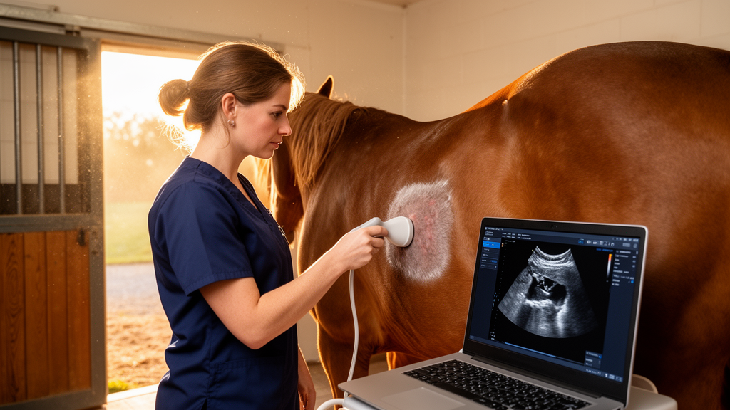
A study published in BMC Veterinary Research now reports a proof-of-concept deep learning tool that can recognize the standardized ultrasound windows of the FLASH protocol — Fast Localised Abdominal Sonography of Horses — directly from ultrasound images, a first step toward computer-assisted support for this emergency examination.
FLASH is a focused point-of-care ultrasound protocol used in most equine hospitals to rapidly assess horses with colic and other emergencies. It examines a thoracic window plus six abdominal windows on both sides of the horse, evaluating intestinal contents, wall thickness, distension, motility and free fluid, as well as the presence and size of the kidney and stomach. Previous work has shown the technique to be more accurate for detecting lesions requiring surgical intervention than transrectal palpation, which is why rapid and accurate window recognition matters so much in the clinic.
The new study, a retrospective-prospective proof-of-concept investigation conducted at Ludwig-Maximilians-University in Munich, set out to develop software that identifies FLASH-specific windows and recognizes the ultrasonographic organ patterns within them. Rather than aiming at autonomous diagnosis, the authors framed the tool primarily as an educational aid: a supplement to conventional teaching that would let veterinary students practice basic probe orientation and standardized window recognition before scanning live horses, in a field where ultrasound teaching is underrepresented despite being considered a first-day competency.
The dataset came from just six healthy horses owned by the university — a Haflinger stallion, a Haflinger mare, a Warmblood mare, two Standardbred mares and a Thoroughbred mare, with a median age of 19 years. Examinations were performed by one resident and one board-certified diplomate of the European College of Equine Internal Medicine using a GE Logiq Vivid E95 machine with a convex array transducer at 2.8 MHz, following the FLASH protocol described by Busoni and colleagues in 2011. Cine loops recorded during repeated examinations were decomposed into individual frames, which two ECEIM board-certified examiners assigned to their respective windows.
The engineering behind the classifier reflects careful attention to a subtle but critical pitfall in medical image artificial intelligence: data leakage. Because frames extracted from a single cine loop are highly similar, splitting them across training and test sets would inflate performance estimates. The team therefore partitioned the dataset at the examination level, keeping every frame from one acquisition within a single partition. Frames were preprocessed to strip textual annotations and everything outside the ultrasound cone, filtered by a black-level threshold, and manually quality-controlled to remove empty frames, motion artifacts and images with incomplete ultrasound sectors.
Not every window survived this pipeline. FLASH windows 4 and 6, the left and right middle third of the abdomen, were substantially underrepresented in the data and showed considerable anatomical overlap — in healthy horses both predominantly display large intestinal structures with nearly identical sonographic appearance. Despite repeated optimization attempts, reliable classification of these two windows proved impossible, and they were excluded from the final model. From an initial 23,454 images across the remaining five windows, 211 images were removed automatically and 3,243 more after manual review, leaving 20,000 images, or 85.27 percent of the original dataset. A cap of 99 frames per examination limited overrepresentation of individual scans.
The final model used EfficientNet-B0, a convolutional neural network architecture pretrained on ImageNet, as its backbone. Hyperparameters were tuned with the Optuna framework over ten trials using three-fold cross-validation, with mean validation accuracy as the optimization objective, and data augmentations such as flipping and color jittering were dropped after failing to improve performance. On an independent test set of 6,119 images that the model had never seen, the classifier achieved an overall accuracy of 80.8 percent, with macro-averaged precision, recall and F1-score of 0.808, 0.787 and 0.767 respectively.
Performance varied sharply between windows. The duodenal window was the star: every single test image was correctly classified, a recall of 1.00, with a precision of 0.884 and an F1-score of 0.938. The nephrosplenic window also performed strongly, with a precision of 0.965, though about 23 percent of its images were misclassified as the gastric window — an asymmetric confusion, since only 2 percent of gastric images went the other way. The gastric window itself landed in balanced middle territory with a precision of 0.820 and recall of 0.781. The ventral window achieved high recall of 0.844 but low precision of 0.407, signaling frequent false positives, while 16 percent of ventral images were predicted as duodenal.
The most dramatic failure involved the thoracic window. Only 55 percent of thoracic images were correctly identified, and 45 percent were misclassified as the ventral window — nearly half the class swept into its abdominal neighbor. Attention heat maps generated with Grad-CAM offered a possible clue: in some thoracic images the model’s attention shifted toward the image border rather than the underlying anatomy, suggesting reliance on non-anatomical image features. The authors note that the model was trained on isolated frames rather than video sequences, so it could not use dynamic information such as respiratory motion or probe movement, and that images lacking clearly visible pleural landmarks may simply resemble ventral abdominal views dominated by soft tissue patterns and gas artifacts.
The study’s limitations are stated candidly and they matter for how the results should be read. Ground truth itself was fuzzy, because FLASH windows are operator-defined regions with partially overlapping anatomical content rather than strictly separated compartments, and selection bias entered through the examiners’ interpretation of windows and their definition of normal physiological images. The dataset was not homogeneously dispersed across windows, and — most importantly — it derived from only six horses, meaning different examinations from the same animal could appear in both training and test data, so the model may have learned horse-specific characteristics and the reported performance may be overestimated. The authors explicitly call for validation on independent datasets from previously unseen horses, larger and more balanced multi-operator datasets, external validation and video-based analysis.
Even with those caveats, the authors argue the work demonstrates feasibility rather than clinical readiness, and their conclusion is explicit that classification performance is not yet sufficient for routine clinical or educational use. Misclassifications concentrated between anatomically related and adjacent windows, which the team interprets as evidence that the model was grappling with genuinely ambiguous anatomy rather than random noise — a problem amplified in healthy horses, where pathological changes such as distension or altered wall thickness that make regional differences diagnostically meaningful are absent. Future incorporation of organ-level annotations and probe position information may improve discrimination between anatomically similar windows.
The longer-term vision extends into the teaching laboratory. The researchers propose developing a virtual reality ultrasound dummy built on this recognition tool, allowing students and inexperienced veterinarians to practice window recognition without repeated examinations on live horses — an approach the authors link to the 3Rs framework of replacement, reduction and refinement, easing the burden on teaching horses while building skills earlier. They are careful to position any such system as decision support rather than a replacement for clinical expertise: ultrasound findings in colic must always be integrated with physical examination and patient history, and the algorithm’s role is to assist standardization and interpretation, especially for less experienced operators, not to deliver autonomous diagnostic conclusions.
Subject of Research: Deep learning classification of equine FLASH ultrasound windows in healthy horses
Article Title: Deep learning-based differentiation of FLASH ultrasound windows
Article References: Köninger, L. M., Müller, T., Gesell-May, S., Cavalleri, J.-M. V., Kau-Strebinger, S., & May, A. (2026). Deep learning-based differentiation of FLASH ultrasound windows. BMC Veterinary Research, 22(1), Article 625. https://doi.org/10.1186/s12917-026-06021-6
Image Credits: AI Generated
DOI: 10.1186/s12917-026-06021-6
Keywords: deep learning, FLASH protocol, equine colic, veterinary ultrasound, EfficientNet-B0, image classification, point-of-care ultrasound, veterinary education, proof-of-concept study, horses, artificial intelligence, diagnostic imaging
News Source: William Thompson. (October 29, 2026). AI Learns to Read Equine Emergency Ultrasound Windows in Proof-of-Concept Study. Scienmag.