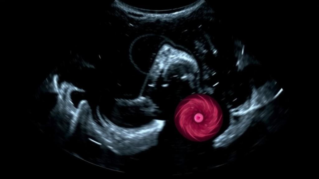
Breast ultrasound is one of the most widely used tools for investigating suspicious lumps, prized for being radiation-free, inexpensive and portable. Yet teaching artificial intelligence to read these scans reliably has long been hampered by a stubborn bottleneck: a shortage of expertly annotated images. Now, researchers at Brno University of Technology in the Czech Republic have reported a framework that tackles the problem head-on by teaching a neural network to manufacture the very training data it needs. The study, published in BMC Medical Imaging by Ali Hamza and Martin Mezl, introduces UltraGANet, a system that pairs a conditional generative adversarial network with a lightweight convolutional classifier to sort breast ultrasound images into benign, malignant and normal categories.
The core insight behind the work is deceptively simple. Deep learning models are voracious consumers of labeled data, and medical imaging is notoriously stingy with it. Annotating ultrasound scans requires trained radiologists, patient consent and institutional infrastructure, all of which constrain dataset size. Small datasets, in turn, leave models vulnerable to overfitting, the phenomenon in which a network memorizes the quirks of its training examples rather than learning the underlying patterns that generalize to new patients. Data augmentation, the practice of artificially expanding a training set, has therefore become a central strategy in medical machine learning, and generative adversarial networks represent one of its most powerful expressions.
A generative adversarial network pits two neural networks against each other in a competitive game. The generator produces synthetic images from random noise, while the discriminator tries to distinguish those fakes from real examples. As training proceeds, the generator grows increasingly adept at producing images that fool the discriminator, effectively learning the statistical structure of the real data. In the conditional variant used by the Brno team, the generator receives an additional input specifying the desired class, allowing it to produce images on demand that carry the visual characteristics of benign tumors, malignant tumors or normal breast tissue. This class-conditional control is what makes the approach practical for building a balanced, class-specific training corpus.
Hamza and Mezl developed their conditional GAN to generate class-specific grayscale breast ultrasound images and then combined these synthetic samples with real images from the BUSI dataset, a publicly available collection of breast ultrasound scans, to construct an augmented training set. The classifier component, UltraGANet, was deliberately kept lightweight, a design choice with practical consequences. Compact convolutional networks demand less memory and computational power than their giant counterparts, which matters enormously in clinics and hospitals that lack access to high-end graphics hardware. The authors position the framework explicitly as a candidate for data-scarce and resource-limited settings, where both annotated images and computing infrastructure are in short supply.
Evaluation was carried out with a rigor that lends the results credibility. The team used five-fold stratified cross-validation, a technique in which the data is divided into five partitions and the model is trained and tested five separate times, each time holding out a different partition, with stratification preserving the class balance across splits. They also assessed the model on an independent test set of 146 real ultrasound images that the classifier had never seen during training. This two-pronged evaluation matters because cross-validation alone can flatter a model when synthetic data leaks across folds; the independent real-image test provides a more honest estimate of clinical performance.
The numbers tell a consistent story. UltraGANet achieved a mean cross-validation accuracy of 0.914, with a standard deviation of 0.012, indicating stable performance across the five folds. On the independent test set of real images, accuracy reached 0.904. Perhaps most significantly for clinical utility, the model’s recall on malignant tumors hit 0.92, meaning it correctly identified 92 percent of the cancerous cases in the test set. In cancer screening and diagnosis, recall on the malignant class is arguably the single most important metric, because a missed malignancy carries far graver consequences than a false alarm that triggers follow-up imaging. Precision and F1-scores, which balance precision against recall, were generally consistent across the three classes, suggesting the model did not simply excel on one category at the expense of the others.
The authors also interrogated the quality of the synthetic images themselves, a crucial step because poor-quality fakes can teach a classifier the wrong lessons. Qualitative inspection and quantitative evaluation suggested that the generated images retained class-relevant ultrasound characteristics and showed moderate structural similarity to real scans. Metrics of this kind, such as structural similarity measures, compare synthetic images against real ones to verify that the generator has captured genuine anatomical and textural features rather than producing plausible-looking noise. The moderate degree of similarity reported here reflects a common trade-off in generative modeling: images that are too similar to real training examples risk simple memorization, while images that are too different may not carry useful signal.
What makes this study noteworthy in the broader landscape of AI-assisted diagnostics is the combination of generative augmentation with an intentionally modest classifier. Much of the headline-grabbing work in medical imaging relies on massive pretrained architectures with hundreds of millions of parameters, which can be impractical to deploy in the very settings where ultrasound is most valuable. Ultrasound machines are often the first, and sometimes the only, imaging modality available in rural clinics and lower-resource health systems. A framework that can run on modest hardware while still achieving above-ninety-percent accuracy on real images speaks directly to that deployment reality, and the authors report that the cGAN-based augmentation improved classification performance compared with training on real images alone, particularly for malignant tumor detection.
Caution remains warranted before such systems reach the clinic. The study used a publicly available dataset rather than newly collected patient data, and the authors note that no new patient data was gathered, so the model has yet to face the messier variability of multi-center, multi-vendor ultrasound machines, different transducer settings and diverse patient populations. The independent test set, while real, comprised 146 images, a modest sample by clinical validation standards. The article itself was shared early as an accepted manuscript subject to further edits, carrying a permanent DOI and citable status ahead of the final version of record. These are normal caveats in the translation of machine learning research into medical practice, but they underline that a promising cross-validation score is a beginning, not an endpoint.
Even so, the trajectory of the work is emblematic of where medical AI is heading. Rather than waiting for ever-larger annotated datasets that may never materialize, researchers are increasingly asking generative models to fill the gap, synthesizing the rare, the underrepresented and the expensive-to-collect. In breast imaging, where early detection dramatically improves outcomes and where benign findings vastly outnumber malignant ones, the ability to generate balanced, class-specific training data could help classifiers learn the subtle textural signatures of malignancy without requiring thousands of additional biopsied scans. Hamza and Mezl’s UltraGANet offers a compact, open and reproducible demonstration that this strategy can deliver measurable gains today, and it points toward a future in which the training data for diagnostic AI is, in part, dreamed up by the machines themselves.
Subject of Research: Conditional GAN-based data augmentation for deep learning classification of breast tumors in ultrasound imaging
Article Title: UltraGANet: a class-conditional GAN framework for breast tumor classification in ultrasound imaging
Article References: Hamza, A., & Mezl, M. (2026). UltraGANet: a class-conditional GAN framework for breast tumor classification in ultrasound imaging. BMC Medical Imaging. https://doi.org/10.1186/s12880-026-02841-y
Image Credits: AI Generated
DOI: 10.1186/s12880-026-02841-y
Keywords: breast cancer, ultrasound imaging, generative adversarial networks, conditional GAN, deep learning, convolutional neural network, medical image analysis, data augmentation, tumor classification, BUSI dataset, computer-aided diagnosis, BMC Medical Imaging
News Source: Ophelia Keating. (October 13, 2026). AI Learns to Fake Ultrasound Images to Spot Breast Tumors More Accurately. Scienmag.