


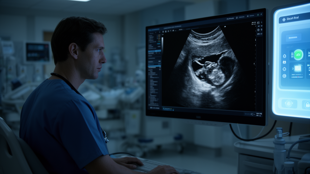
When a patient arrives at an emergency department with severe abdominal pain, every minute matters. One of the most common first-line tests is an ultrasound of the right upper quadrant of the abdomen, an imaging study that visualizes the liver, gallbladder, right kidney and part of the pancreas. Now, researchers at the University of Colorado Anschutz School of Medicine have built an artificial intelligence agent that can read these scans, diagnose common conditions affecting those organs, and draft clinical reports, potentially shaving precious minutes off the path to treatment.
The new system, described in a study published in Nature Communications, is what the researchers call a vision-language AI agent. Unlike a model that looks at images alone, this tool blends the visual data from the ultrasound with written clinical information from the laboratory, such as blood test results. The combination allows the agent to interpret the scan in the context of the patient’s broader clinical picture, diagnose common health conditions affecting the organs in view, and suggest logical next steps for care.
Harrison Bai, MD, MS, a visiting associate professor in the Department of Radiology at CU Anschutz and senior author of the paper, stresses that the agent is designed to work alongside a trained radiologist rather than instead of one. The system generates reports that the radiologist then reads and evaluates. “The goal is not to replace the physician. We’re not trying to build an AI radiologist,” Bai says. “But if our AI agent can write up preliminary reports, it saves radiologists time.”
Speed matters most in emergencies. Patients with acute gallbladder inflammation, a condition known as cholecystitis, need emergency surgery right away, and the AI tool can rapidly flag patients who may need urgent operations so they can reach the operating table as quickly as possible. According to Bai, the vision-language model can generate reports quickly, which is especially crucial when a patient is experiencing a medical emergency, allowing diagnosis and surgery to happen sooner.
Right upper quadrant ultrasounds are often doctors’ top choice for diagnosing problems involving the liver or the biliary system. Physicians typically order them when patients have pain in the right upper abdomen or certain abnormal laboratory results, such as elevated liver enzymes or bilirubin. The scans are painless and non-invasive, and they can help identify a range of conditions including gallstones, fatty liver, liver cirrhosis, hepatitis, kidney stones and pancreatitis, and may even potentially find cysts or tumors in these organs.
The push for automation comes against a backdrop of mounting pressure on the radiology workforce. Many fields of medicine face specialist shortages, but radiologists face extra strain from an aging U.S. population and rising demand for medical imaging. Patient loads are increasing while doctors are asked to accomplish more with fewer resources, and as of 2022 radiologists were leaving the field at twice the rate they were in 2014. “We’re trying to address the increasing workload. It takes time to write reports on imaging results. There aren’t enough people or radiologists to do the work, and there’s decreasing reimbursement by Medicare insurance,” Bai says. “If you tell radiologists that an AI tool can save them time, suddenly, people are much more likely to use the technology.”
Interpreting these scans is also technically difficult. The people who read right upper quadrant ultrasounds have varying levels of skill and experience, and the images themselves can contain significant visual noise and artifacts that less-trained interpreters may not recognize. That means diagnostic accuracy often depends on who is reading the study. The researchers argue that incorporating a vision-language model could help standardize interpretation, improve accuracy, and make results more consistent across different readers.
The team designed the model to read right upper quadrant ultrasounds, scan them for 16 common diagnoses, estimate each patient’s prognosis, and automatically generate reports. To test it, the researchers used a dataset of roughly 11,000 ultrasounds drawn from Stanford University, the University of Colorado and Johns Hopkins University, then manually reviewed 100 of the AI-generated reports for accuracy. In that sample, the model identified the correct diagnoses in each ultrasound 99.6% of the time. It did make some mistakes in the overall reports, but the radiologists who reviewed and edited the reports found those errors easy to catch.
The researchers also ran a blinded evaluation in which a panel of experts tried to distinguish reports written by the AI from reports written by radiologists themselves. The experts could pick out unedited, AI-written reports most of the time, but they could not reliably tell radiologist-edited AI reports apart from reports written entirely by radiologists. In the paper, the team describes how the model should operate as a robust, clinically integrated assistant that integrates different types of information and helps with clinical decision-making without replacing the expertise a radiologist provides.
Before the technology can be widely adopted, the researchers caution that its performance must be evaluated in real-world emergency care, and that it will be critical to study how physician-AI collaboration affects diagnostic accuracy, efficiency and patient well-being. Bai does not expect AI to replace the need for human supervision in medicine, and he acknowledges it will take time for doctors to adapt, but he believes the technology could change the course of medicine for the better. “I can see this technology playing a huge role in how medicine is practiced,” he says. “It will involve a lot of change and learning about new things. But I’m super excited. I want to see these developments not just happening in my lab, but also incorporated into daily practice to help patients and doctors.”
Subject of Research: A vision-language AI agent for interpreting right upper quadrant ultrasounds and supporting radiologist decision-making
Article Title: AI tool could help radiologists interpret ultrasounds faster and more accurately
Article References: AI tool could help radiologists interpret ultrasounds faster and more accurately. Original publication (Bibliographic details unavailable.)
Image Credits: AI Generated
DOI: Not provided
Keywords: artificial intelligence, radiology, ultrasound, vision-language model, emergency medicine, gallbladder, cholecystitis, Nature Communications, diagnostic accuracy, radiologist workload, CU Anschutz, clinical decision support
News Source: Ophelia Keating. (October 30, 2026). AI agent reads emergency abdominal ultrasounds with 99.6% diagnostic accuracy in study. Scienmag.



