
Hepatocellular carcinoma (HCC) is a pressing global health crisis that ranks as the sixth most prevalent cancer while being the third leading cause of cancer-related mortality. The substantial burden posed by HCC is compounded by existing treatment challenges that stem from the disease’s often late diagnosis and the inherent complexities of liver anatomy. Traditional management strategies frequently employ hepatectomy, which is a surgical resection of the liver. For surgeons, the accurate detection and complete excision of tumor tissue are crucial for attaining positive patient outcomes. Unfortunately, conventional imaging methods fall short when it comes to providing real-time guidance during surgical interventions. This gap in technology has prompted the healthcare community to investigate fluorescence-guided surgery (FGS) as an innovative approach to enhance surgical precision and decision-making.
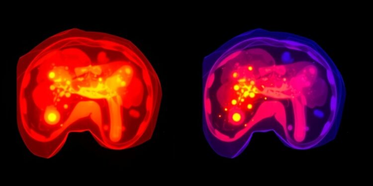
Fluorescence-guided surgery utilizes specific fluorescent dyes to illuminate tumor tissues during surgical procedures, leading to enhanced visualization for the operating surgeon. Among the array of fluorescent agents, indocyanine green (ICG) has garnered significant attention and is frequently employed in FGS. ICG’s molecular structure allows it to bind with plasma proteins, which facilitates its selective absorption by hepatocytes. The unique metabolic pathways of HCC cells reveal a propensity for ICG accumulation due to disrupted biliary excretion mechanisms. This characteristic makes it possible to visualize cancerous tissues during surgery. Yet, despite the utility of ICG, several limitations persist, including its insufficient specificity in distinguishing cancerous from healthy liver tissue, as well as challenges in imaging deeper lesions within the liver anatomy.
Recognizing the drawbacks presented by ICG, researchers are embarking on a journey to develop novel fluorescent probes that may offer more precise and robust tumor detection capabilities. These innovative compounds encompass a variety of mechanisms, including the use of enzymes, reactive oxygen species (ROS), reactive sulfur species (RSS), and pH-sensitive agents. Among these cutting-edge probes, aggregation-induced emission (AIE) probes stand out due to their unique optical properties. AIE probes exhibit minimal fluorescence in dilute solutions; however, they become highly fluorescent when aggregated, which is ideal for applications such as real-time surgical navigation. Several advantages characterize AIE probes: superior biocompatibility ensures minimal toxicity to surrounding tissues, high photostability allows for extended visualization, and reduced background signal interference enhances the clarity of tumor detection.
Clinical endeavors utilizing ICG for fluorescence guidance have demonstrated promising outcomes, facilitating the delineation of tumor borders and enabling the identification of small lesions that would otherwise remain undetectable. Yet, the clinical application of FGS is not devoid of challenges. A significant hurdle arises when attempting to visualize deeper structures due to the limited penetration of near-infrared light, which is crucial for optimal ICG imaging. Moreover, the uptake and distribution of ICG can be significantly influenced by underlying conditions such as liver cirrhosis or variations in hepatic blood flow, all of which complicate its effective use in heterogeneous patient populations. Additionally, benign liver conditions can generate fluorescence signals similar to those from malignant tissue, thereby adding a layer of complexity in accurately differentiating between the two.
As the future of fluorescence-guided surgical techniques evolves, trailed by groundbreaking studies, the focus is shifting toward developing the next generation of fluorescent probes with enhanced imaging characteristics. A considerable aspect of this research focuses on improving tissue penetration and targeting specificity, particularly through the exploration of near-infrared fluorophores operating within the 1,000-1,700 nm range. This spectral region promises to provide greater imaging depth while maintaining high spatial resolution. Furthermore, there is a growing interest in the integration of fluorescence imaging with complementary imaging techniques such as computed tomography (CT) and magnetic resonance imaging (MRI). Such multimodal approaches could significantly improve diagnostic accuracy and augment surgical outcomes, ultimately benefiting the surgical management of HCC.
The ultimate aspiration in the realm of fluorescence-guided surgery is to equip surgeons with cutting-edge tools that enable them to perform more precise tumor resections, a feat that may contribute to reducing recurrence rates and enhancing overall survival for patients afflicted with hepatocellular carcinoma. Researchers and clinicians are constantly working to refine existing technologies and evaluate new applications, with several promising agents currently under preclinical and clinical evaluation. As the body of research surrounding fluorescent probes expands, the prospects they present for revolutionizing oncologic surgery and alleviating the burden of liver cancer become increasingly tangible.
There is a wealth of academic analysis surrounding the integration of fluorescence-guided techniques into surgical protocols that reflects the potential transformative impact of these advances on patient outcomes in liver cancer. Collaborating institutions and researchers from various domains, including surgical oncology, biomedical engineering, and molecular biology, are beginning to bridge gaps and work toward a comprehensive understanding of how fluorescence-guided technologies can support the management of HCC. This collaborative effort underscores an optimistic future for surgical oncology, fostering an environment ripe for innovation and enhanced therapeutic strategies. The convergence of science and clinical practice embodies the essence of progress in achieving improved patient care through the application of advanced imaging and guiding technologies.
As the clinical community eagerly anticipates breakthroughs in this domain, the message is clear: fluorescence-guided surgery holds incredible promise in the fight against hepatocellular carcinoma. Through careful study, innovative probe development, and multidisciplinary research collaborations, there lies an exciting opportunity to redefine surgical management and improve results for patients worldwide impacted by liver cancer. The ongoing evolution of this field will continue to provide insights and advancements that pave the way for significant developments in surgical oncology and its approach to liver disease.
Subject of Research: Fluorescence-guided surgery for hepatocellular carcinoma
Article Title: Fluorescence-guided Surgery for Hepatocellular Carcinoma: From Clinical Practice to Laboratories
News Publication Date: 2-Jan-2025
Web References: Journal of Clinical and Translational Hepatology
References: N/A
Image Credits: N/A
Keywords: Hepatocellular carcinoma, liver tumors, fluorescence microscopy, cancer treatments, clinical research
Tags: bridging clinical practice and researchcancer-related mortality statisticschallenges in liver cancer managementenhancing surgical outcomes in HCCfluorescence-guided surgery techniqueshepatocellular carcinoma treatmenthepatocyte fluorescence uptakeindocyanine green in surgeryinnovative approaches to cancer surgeryreal-time imaging for HCCsurgical precision in liver cancertumor tissue visualization methods
