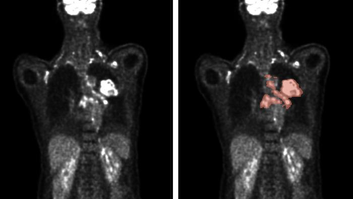
Imaging techniques play a pivotal role in diagnosing cancer, a disease characterized by the uncontrolled growth of abnormal cells. Among these techniques, positron emission tomography (PET) and computed tomography (CT) stand out due to their complementary capabilities. PET is primarily focused on metabolic activity, utilizing radioactively labeled glucose, especially fluorine-18-deoxyglucose (FDG), to visualize the heightened metabolic rates inherent in malignant tumors. In contrast, CT leverages X-ray technology to capture detailed anatomical images of the body, providing critical insights into the location and structure of tumors. Both modalities, when used in conjunction, create a comprehensive view that is essential for effective diagnosis and therapy selection.
Cancer patients often present with a multitude of lesions, making the accurate assessment of size and number paramount for effective treatment planning. The sheer volume of lesions can be overwhelming, leading to a labor-intensive process where clinicians manually identify and quantify these tumors from 2D images. This manual approach not only demands an immense amount of time but is also prone to human error, adding layers of complexity to oncological evaluations. To mitigate these issues, researchers are turning to automated evaluation systems that harness advanced algorithms to enhance accuracy and reduce workload.
Professor Rainer Stiefelhagen, who leads the Computer Vision for Human-Computer Interaction Lab (cv:hci) at the Karlsruhe Institute of Technology (KIT), advocates for automated methods, emphasizing their potential to revolutionize tumor assessment. He points out that an algorithm can drastically cut down the time required for evaluation while simultaneously improving diagnostic precision. This sentiment is echoed by a growing body of evidence that suggests automated systems can surpass traditional manual methods in both speed and accuracy.
In the realm of automated PET and CT imaging, the autoPET challenge serves as a highlight, showcasing the capabilities of modern machine learning techniques. In 2022, Stiefelhagen, along with doctoral student Zdravko Marinov and collaborators from the IKIM Institute for Artificial Intelligence in Medicine, participated in this international competition. The event brought together 27 teams with 359 participants worldwide, all striving to create algorithms capable of automatically segmenting metabolically active tumor lesions in whole-body PET/CT scans.
The crux of the challenge was to employ deep learning methods, which are designed to recognize patterns in extensive datasets through multi-layered artificial neural networks. These algorithms were tasked with analyzing large, annotated PET/CT datasets, significantly enhancing their ability to learn and adapt to complex imaging data. As a result, the competition offered an invaluable opportunity for teams to benchmark their approaches against others on a global scale, ultimately leading to breakthroughs in automated analysis.
After rigorous testing, the results indicated that an ensemble of top-performing algorithms outshone their individual counterparts, demonstrating efficiency and precision in tumor detection. The ensemble approach capitalizes on the strengths of multiple algorithms, compensating for their individual weaknesses to deliver robust performance. Stiefelhagen explains that while data quality and quantity significantly influence algorithm performance, the design and execution of post-processing decisions are critical in achieving optimal results. This intricate balancing act is essential for ensuring that algorithms can make accurate assessments in a clinical environment.
Despite the strides made in automated imaging analysis, further research is vital to enhance these algorithms’ resilience against external factors. As researchers aim for a future where the analysis of PET and CT images is fully automated, they acknowledge that clinical deployment will necessitate not just precision but also adaptability in various real-world scenarios.
The landscape of medical imaging continues to evolve, with automated solutions emerging as a cornerstone in oncological diagnostics. The implications of successfully deploying these systems are profound, potentially leading to earlier detection of cancers, more personalized treatment plans, and ultimately improved patient outcomes. Stiefelhagen and his colleagues are at the forefront of this technological revolution, with their recently published research in the esteemed journal Nature Machine Intelligence paving the way for future advancements in medical imaging.
The autoPET challenge not only demonstrates the potential of automated systems but also underscores the importance of collaboration across disciplines and institutions. By combining expertise in computer science, medicine, and imaging, researchers can foster innovations that hold the promise of transforming cancer diagnostics and treatment. As the medical community embraces these cutting-edge technologies, the journey toward enhanced diagnostic accuracy continues to gather momentum.
In conclusion, the integration of automated analysis in PET and CT imaging marks a significant step forward in oncological care. By harnessing the power of machine learning and deep learning algorithms, researchers are unlocking new avenues for improving cancer diagnosis and treatment planning. As this field progresses, ongoing collaboration and innovation will be essential to realizing the full potential of automated imaging solutions. In the quest to provide better care for cancer patients, the fusion of technology and medicine remains a beacon of hope.
Subject of Research: People
Article Title: Results from the autoPET challenge on fully automated lesion segmentation in oncologic PET/CT imaging.
News Publication Date: 30-Oct-2024
Web References: Nature Machine Intelligence DOI
References: Sergios Gatidis, et al., Results from the autoPET challenge on fully automated lesion segmentation in oncologic PET/CT imaging. Nature Machine Intelligence, 2024. DOI: 10.1038/s42256-024-00912-9
Image Credits: Credit: Gatidis S, Kuestner T, 2022.
Keywords: medical imaging, cancer diagnosis, PET, CT, machine learning, deep learning, automated segmentation, oncology.
