
In a groundbreaking study, researchers from East China University of Science and Technology have developed activatable organic phosphorescent probes that promise substantial advancements in bioimaging and biosensing technologies. Led by Professor Xiang Ma and Dr. Yang Li, this pioneering research explores the synthesis and application of red/near-infrared room temperature phosphorescence probes, engineered through a supramolecular assembly technique involving macrocyclic compounds and guest molecules.
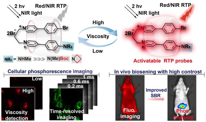
The main focus of their research was on an innovatively designed L1C probe, which exhibited remarkable properties that are crucial for cellular imaging applications. Integrating secondary amino groups, the probe was optimized to enhance its phosphorescent qualities when exposed to varying levels of solution viscosity. The researchers found that introducing specific molecular modifications, including the Boc group and nitrogen heterocyclic butyl moieties, significantly improved the emissive characteristics of these optical probes. This study leverages host-guest chemistry to fully exploit the exciting potentials of organic phosphorescence.
As the viscosity of the surrounding solution was altered, the L1C probe demonstrated not only an increase in phosphorescence intensity but also an elongation of its lifetime. This remarkable feature allows researchers to decipher complex biological interactions and processes at unprecedented resolution. The implications of these structural and performance enhancements are vast, as they open new avenues for time-resolved imaging—essential in biological studies that require precise monitoring of cellular dynamics.
Moreover, the resultant L1C probe was evaluated for its biocompatibility and specificity in targeting lysosomes, marking a significant step toward utilizing phosphorescent materials for real-time imaging in living organisms. Proven to exhibit targeted delivery and resilience in biological environments, L1C has the potential to transform the landscape of in vivo imaging techniques. Such advancements not only amplify the capabilities of bioimaging but also lay the groundwork for better therapeutic monitoring in clinical settings.
The research team employed sophisticated methods to validate their peptide-based probes through various biological imaging modalities, including two-photon microscopy. These approaches facilitated the observation of cellular viscosity fluctuations, an important indicator of physiological and pathological changes in living organisms. The ability to monitor such dynamic parameters in real-time grants scientists a powerful tool for understanding disease mechanisms and cellular responses to treatment.
Interestingly, the innovation did not stop at in vitro applications. In vivo experiments showcased the efficacy of the L1C probe to visualize viscosity alterations in an inflammation mouse model. The results revealed a remarkably high signal-to-background ratio, exceeding 80, underscoring the probe’s ability to deliver reliable data amid complex biological systems. This capability is essential for accurate biosensing and diagnostic applications, paving the way for future studies into various diseases, including cancer and inflammatory conditions.
Dr. Yang Li articulated the pivotal role these activatable phosphorescent probes could play in advancing biomedical research, stating that their development could revolutionize how organic room-temperature phosphorescent materials are utilized in imaging. As imaging technology evolves, these probes are anticipated to integrate seamlessly with emerging methodologies, enhancing diagnostic precision and enabling intricate insights into biological processes.
The significance of this research extends beyond merely improving existing imaging technologies. The researchers propose that their activatable red/near-infrared phosphorescent probes can reflect the complexities of dynamic biological microenvironments with high sensitivity. This real-time capability could dramatically reshape how scientists explore molecular markers and pathological alterations within cells, delivering breakthrough insights into disease progression.
Through their work, Li and Ma have not only contributed to the field of materials science but also enriched our comprehension of biological systems. Their findings represent a critical evolution in phosphorescence imaging, signifying a step toward more accurate diagnosis and responsive treatment protocols. The ongoing advancements in these probe technologies exemplify the intersecting paths of chemistry, biology, and medicine.
In conclusion, the development of activatable organic phosphorescent probes heralds a new era in biological imaging, with profound implications for future research and clinical applications. As researchers continue to explore the multifaceted roles of these probes in various biological contexts, the potential for more detailed and accurate imaging shall only expand, helping to illuminate the intricacies of life at a fundamental level.
As the study paves the way for innovations in phosphorescence, it stands as a testament to the power of interdisciplinary research and collaboration, highlighting the remarkable progress being made in the realm of biophysics and bioengineering.
Subject of Research: Activatable Organic Phosphorescent Probes
Article Title: Activatable red/near-infrared aqueous organic phosphorescence probes for improved time-resolved bioimaging
News Publication Date: October 2023
Web References: Science China Press
References: National Science Review
Image Credits: Credit: ©Science China Press
Keywords: organic phosphorescent probes, bioimaging, fluorescence, viscosity detection, inflammation monitoring, cellular imaging, supramolecular assembly, lysosome targeting, real-time imaging, advanced diagnostics.
