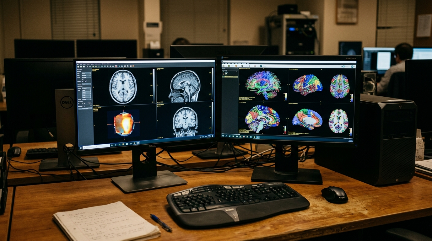
A deep learning architecture combining structural magnetic resonance imaging and resting-state functional brain scans has established a new benchmark in classifying autism spectrum disorder across diverse medical imaging centers. Developed to overcome individual biological heterogeneity and hardware variation across clinical scanners, the cross-attention-guided subject-adaptive graph network, designated CAS-GNN, achieved an average accuracy of 78.75 percent across five-fold cross-validation and 79.25 percent on completely held-out independent test subjects from the international Autism Brain Imaging Data Exchange repository. The computational framework simultaneously resolved an area under the receiver operating characteristic curve of 0.804 while yielding an independent test precision of 78.57 percent, a recall of 81.48 percent, an F1 score of 80.00 percent, and a specificity of 76.92 percent. By systematically anchoring dynamic functional connectivity to persistent anatomical structures, the study demonstrates that multimodal representations can reliably separate individuals with autism from typical controls while providing interpretable insights into altered cortical networks.
Autism spectrum disorder is a complex neurodevelopmental condition characterized by social communication difficulties and repetitive behaviors. For years, neuroscientists have investigated brain imaging as a promising diagnostic aid, yet clinical translation has been hindered by substantial structural and functional variation among autistic individuals. Prior computational investigations predominantly relied on unimodal features, examining anatomical metrics from structural magnetic resonance imaging or connectivity metrics from resting-state functional scans in isolation. These single-modality methodologies inevitably provide an incomplete view of the intricate neural mechanisms driving the disorder. When researchers attempted to fuse structural and functional data through simple feature concatenation or global summation, disparate feature scales, differing noise patterns, and mismatched data distributions prevented algorithms from leveraging the true strengths of each modality. Compounding these analytical difficulties, differing scanning hardware, head coils, and acquisition protocols across imaging laboratories introduced significant distributional shifts that reduced generalization across multicenter datasets and compromised clinical utility.
To construct a robust diagnostic pipeline, the researchers examined multicenter neuroimaging cohorts from the Autism Brain Imaging Data Exchange repository, drawing primary experimental data from New York University, UCLA, the University of Utah, and the University of Michigan. Participant selection enforced strict quality control criteria, requiring the concurrent availability of high-quality structural and functional scans, valid diagnostic labels, and head movement constrained to no more than 2.5 millimeters of displacement and 2.5 degrees of rotation. Functional preprocessing discarded the initial five volumes for signal stabilization, corrected slice acquisition timing, aligned head motion, and co-registered functional images to structural scans before normalizing into standard stereotaxic space at three-millimeter isotropic resolution. Nuisance signals, including twenty-four motion parameters, white matter, cerebrospinal fluid, and global signal fluctuations, were regressed alongside Jenkinson mean framewise displacement corrections, followed by bandpass filtering between 0.01 and 0.08 Hertz. Structural processing segmented cerebral gray matter, establishing standardized anatomical coordinate systems.
To translate neuroimaging data into unified representations, the investigators applied the Brainnetome Atlas, parcellating the brain into 246 regions of interest. From functional scans, they calculated regional mean time series and computed resting-state functional connectivity matrices through Fisher z-transformed Pearson correlation coefficients. From structural scans, they extracted five morphological parameters: gray matter volume, average cortical thickness, surface area, mean curvature, and folding index. Rather than operating directly on 246 fine-grained regions, which introduces computational overhead and spatial noise, the team projected the regions into forty-eight gyrus-level representations using dedicated fully connected layers. To achieve cross-modal integration, the model deployed an asymmetric cross-attention mechanism wherein forty-eight functional tokens served as queries while structural tokens acted as keys and values. This mathematical formulation calculated scaled dot-product attention weights to ground subject-specific functional connectivity within stable anatomical contexts, producing enriched multimodal representations where morphological structure directly constrained dynamic functional measurements.
Following cross-attention fusion, the forty-eight multimodal tokens entered a Vision Transformer architecture to model global dependencies and long-distance relationships across the brain. Each Transformer layer utilized multi-head self-attention and feed-forward networks to enable comprehensive information exchange between every pair of cortical nodes. Conventional graph neural networks applied to neuroimaging typically rely on fixed, population-averaged graph structures that enforce identical connectivity arrangements across all participants, ignoring critical inter-individual biological differences. To overcome this limitation, the authors designed a subject-adaptive graph learning module. The output tokens from the Vision Transformer were decoupled into shared and site-specific components, after which pairwise cosine similarities were computed between all forty-eight cortical units for each participant. For every node, the framework preserved the top-k highest similarity edges alongside self-connections to establish an individualized directed adjacency matrix, capturing subject-specific brain network interactions that adaptively reflected each participant’s distinct structural and functional topology.
With individualized directed graph structures constructed, the architecture applied Chebyshev graph convolutions to achieve stable spectral domain feature propagation across brain nodes. By employing a K-order Chebyshev polynomial expansion, the Chebyshev convolution aggregated feature representations across multi-hop topological neighborhoods while preventing gradient instability. Because standard Chebyshev convolutions require symmetric adjacency matrices and cannot delineate directional signaling between interacting brain regions, the model subsequently routed the spectral representations through a Graph Attention Network. This graph attention module applied learnable linear transformations and attention parameters to calculate directional relevance weights between each cortical node and its connected topological neighbors. By aggregating features across neighboring nodes using dynamic attention coefficients, the network characterized the specific directional flow of neural communication across cortical gyri. Finally, node representations output by the graph attention layers were aggregated through a graph pooling readout mechanism, forming a graph-level representation evaluated by a fully connected classifier.
To overcome scanner-induced distribution discrepancies that hinder multicenter model generalization, the authors implemented a cross-site alignment strategy incorporating domain adversarial training and two-stage supervised contrastive learning. Decoupled shared features from the Vision Transformer were passed through a gradient reversal layer into a domain classifier, utilizing adversarial learning to render shared features indistinguishable across different scanning sites and forcing the network to extract site-invariant biomarkers. Concurrently, two-stage supervised contrastive learning losses were applied to the shared Transformer tokens and the downstream Graph Attention Network outputs. This contrastive framework pulled representations with identical diagnostic labels closer together in latent space while pushing discordant diagnostic representations apart, dynamically balancing contrastive penalties between intra-site and cross-site positive sample pairs. By neutralizing site-specific technical variance while sharpening diagnostic boundaries, the alignment strategy ensured that the classifier learned genuine pathophysiological patterns rather than scanner-specific artifacts, maintaining robust predictive performance across diverse clinical imaging environments.
Rigorous evaluations demonstrated that CAS-GNN substantially outperformed existing graph architectures and traditional machine learning baselines under identical data partitioning protocols. Compared with a dual-branch graph neural network, CAS-GNN achieved a 7.00 percent improvement in classification accuracy, and it exceeded the accuracy of a bilinear perceptual fusion graph convolutional network by 10.00 percent. Traditional classifiers trained on principal component-reduced features, including Support Vector Machines, Random Forests, and XGBoost, were all outperformed by at least 7.47 percent in accuracy. In leave-one-site-out validation across four major ABIDE centers, the model achieved classification accuracies of 70.30 percent at UCLA, 69.11 percent at USM, 67.44 percent at UM, and 63.59 percent at NYU. In independent testing on three previously unseen sites—SDSU, SBL, and Olin—the framework attained average accuracies between 68.51 percent and 72.54 percent, outperforming ablated models lacking cross-site alignment by 3.80 percent and traditional algorithms by more than ten percentage points. Gender-stratified analyses revealed balanced efficacy, registering 77.50 percent accuracy in females and 79.56 percent in males without statistically significant difference.
Ablation experiments demonstrated the vital contribution of each individual component within the CAS-GNN model architecture. Removing the cross-attention module and relying on simple feature concatenation produced a 3.24 percent decrease in classification accuracy, reducing performance to 75.51 percent, with paired statistical testing revealing marginal significance at t = 2.71 and p = 0.053. When multimodal graph construction was replaced with single-modality inputs, accuracy declined by 4.51 percent when utilizing functional connectivity alone and dropped by 5.28 percent when relying solely on structural morphological features. This demonstrates that functional dynamics require anatomical constraints to suppress noise while structural data require functional coupling to capture individual biological differences. Eliminating the cross-site alignment module triggered a 4.39 percent accuracy drop, while removing the subject-adaptive graph learning mechanism caused a statistically significant performance decrease confirmed by paired t-tests at t = 4.67 and p = 0.009. These results confirm that peak diagnostic performance requires the seamless integration of cross-modal attention, subject-adaptive graph modeling, and cross-site adversarial alignment.
Exploratory analyses utilizing eigenvector L2-norm magnitude proxies identified ten primary cortical gyri contributing most heavily to diagnostic classification: the left superior frontal gyrus, right precentral gyrus, right middle temporal gyrus, right inferior temporal gyrus, left fusiform gyrus, right superior parietal lobule, right precuneus, right cingulate gyrus, right medioventral occipital cortex, and left thalamus. The marked predominance of right-hemisphere structures aligns with reported atypical rightward lateralization in autism, reflecting compensatory neural pathways or altered hemispheric specialization. Detailed edge-level evaluations revealed three biologically significant bidirectional connections with group difference magnitudes exceeding 0.2: reciprocal links between the right cingulate gyrus and right medioventral occipital cortex, between the right superior parietal lobule and right cingulate gyrus, and between the right precentral gyrus and right medioventral occipital cortex. Crucially, a support vector regression model confirmed that five specific functional connections significantly predicted clinical Autism Diagnostic Observation Schedule total scores (r = 0.21, p = 0.03, permutation p = 0.005, R2 = 0.067), retaining significance under partial correlation controlling for age and sex (r = 0.28, p = 0.02). Although the authors note sample size constraints at certain sites and the necessity for future multicenter validation on cohorts such as ABIDE-II alongside diffusion imaging or electroencephalography, this interpretable framework establishes an objective, robust foundation for neuroimaging-aided autism diagnosis.
Subject of Research: Multimodal neuroimaging graph learning for autism spectrum disorder classification
Article Title: Cross-attention-guided subject-adaptive graph learning for multimodal autism classification: integrating structural and functional MRI data
Article References: Tang, Y., Yang, C., Xu, Y., Zhang, H., & Xie, H. (2026). Cross-attention-guided subject-adaptive graph learning for multimodal autism classification: integrating structural and functional MRI data. Brain Informatics, 13(1), Article 32. https://doi.org/10.1186/s40708-026-00318-w
Image Credits: AI Generated
DOI: 10.1186/s40708-026-00318-w
Keywords: autism spectrum disorder, neuroimaging, graph neural networks, multimodal MRI, functional connectivity, cross-attention, brain networks, ABIDE repository, Cross-attention-guided, subject-adaptive, graph, learning
News Source: Cassandra Pierce. (November 7, 2026). Adaptive Multimodal Brain Network Model Improves Cross-Site Autism Classification Accuracy. Scienmag.