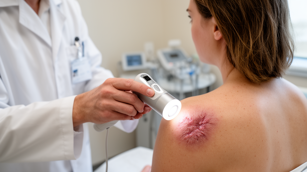
Keloids are among the most stubborn problems in dermatology: raised, fibrotic scars that grow beyond the boundaries of the original wound, often itching, hurting, and continuing to expand for years. For decades, the first-line treatment has been the same — injections of corticosteroid directly into the scar tissue. Yet the response is notoriously unpredictable. Some patients see their keloids flatten and soften over months of treatment, while others endure repeated painful injections with little or no benefit. A new study published in the Archives of Dermatological Research suggests that the answer to which patients will respond may already be visible on the skin surface, captured by an inexpensive, noninvasive imaging tool that dermatologists already use every day: the dermatoscope.
The research, led by Yueming Mu and Yuanyuan Wang of the Department of Dermatology and Venereology at the First Hospital of Jilin University in Changchun, China, set out to answer a deceptively simple question. Could the microscopic surface features of a keloid, observed through a dermatoscope before any treatment begins, predict how well that keloid will respond to intralesional corticosteroid injection over the following year? If so, clinicians could triage patients at the first visit, reserving steroid injections for those likely to benefit and steering others toward alternative therapies sooner, sparing them months of ineffective, uncomfortable treatment.
To investigate, the team enrolled fifty-nine proliferative keloids, all treated with intralesional corticosteroid injection alone, ensuring that any differences in outcome could be attributed to the biology of the lesion rather than to a mix of interventions. Before treatment began, the researchers collected demographic data, clinical characteristics, and Vancouver Scar Scale scores — a validated instrument that grades scars on pliability, height, and vascularity — alongside standardized dermoscopic images of each lesion. The primary endpoint was the treatment response assessed at one-year follow-up, a timeline long enough to capture the slow remodeling processes that determine whether a keloid regresses or resists therapy.
The results revealed just how heterogeneous keloid behavior really is. Of the fifty-nine lesions, twenty — 33.9 percent — showed a good response to treatment, while nineteen, or 32.2 percent, showed no response at all, and another twenty, again 33.9 percent, responded only poorly. In other words, roughly two out of every three keloids in this cohort failed to respond meaningfully to the standard first-line therapy. That statistic alone underscores the clinical frustration that motivates the search for predictive markers: most patients currently begin steroid injections without any way of knowing whether they fall into the fortunate minority or the unresponsive majority.
When the researchers examined the baseline dermoscopic images, two vascular patterns dominated the landscape of these scars. Dotted vessels, appearing as fine red points distributed across the lesion surface, were present in 67.8 percent of keloids, while linear irregular vessels — tortuous, serpentine red lines without a uniform orientation — appeared in 52.5 percent. These patterns are not merely cosmetic curiosities. In dermoscopy, the vascular architecture of a lesion reflects the underlying biology, and in keloids, abundant and visible microvasculature is thought to mirror the intense inflammatory and angiogenic activity that drives fibroblast proliferation and collagen overproduction in the reticular dermis.
The statistical analysis, performed using regression modeling, produced a striking and clinically actionable finding. Keloids with abundant baseline vascularity — whether manifesting as dotted, linear, or branching arborizing vessels — were significantly more likely to respond favorably to corticosteroid injection, with an odds ratio of 0.048 and a 95 percent confidence interval of 0.008 to 0.273, reaching a p-value of 0.001. The direction of the odds ratio reflects how the variable was coded, but the substantive message is unambiguous: a richly vascular dermoscopic appearance marks a keloid that is primed to shrink under steroid therapy. This makes biological sense, because corticosteroids exert part of their anti-scarring effect by suppressing inflammation and reducing vascular supply to the lesion, so a lesion with an active, visible vasculature offers more targets for that mechanism to act upon.
Just as important was the discovery of a marker for treatment failure. The presence of the rosette sign — a distinctive four-quadrant, flower-like white structure seen under polarized dermoscopy — emerged as a strong independent predictor of a poor response, with an odds ratio of 4.500, a 95 percent confidence interval of 1.166 to 17.373, and a p-value of 0.029. Rosettes have been described across a surprising range of conditions in the dermoscopy literature, from basal cell carcinoma to T-cell pseudolymphoma and squamous cell carcinoma, and their optical origin is generally attributed to reflections from stacked, crystalline, or fibrotic structures in the superficial dermis. In the context of keloids, their appearance likely signals a densely remodeled, collagen-rich architecture that is comparatively resistant to the steroid’s anti-inflammatory action — a scar that has already hardened into its final form.
The robustness of these primary findings was reinforced by a sensitivity analysis using Generalized Estimating Equations, a statistical approach that accounts for correlation between observations, which is particularly relevant when multiple lesions from the same patient or repeated measurements are involved. The fact that the vascular-density effect and the rosette-sign effect survived this stricter analysis gives the results a credibility that single-model findings often lack. The study protocol was approved by the Ethics Committee of the First Hospital of Jilin University, and the authors declared no competing interests, with funding provided by the Department of Finance of Jilin Province.
The broader significance of this work lies in its accessibility. Dermoscopy requires no biopsy, no laboratory infrastructure, and no expensive imaging equipment — a handheld dermatoscope costs a fraction of an ultrasound machine or an elastography system, and the examination takes seconds. Other noninvasive technologies have been explored for scar assessment, including shear wave elastography to quantify keloid stiffness and dynamic optical coherence tomography to track vascular changes during laser therapy, and each contributes valuable information. But for the specific clinical decision at hand — should this patient begin a year of steroid injections? — a dermoscopic glance at the vascular pattern may be sufficient. A keloid studded with dotted and linear vessels is a reasonable candidate for first-line injection therapy, while a lesion displaying rosettes might justify an earlier conversation about combination approaches, such as cryotherapy, laser treatment, or surgical excision with adjuvant radiotherapy.
Caveats remain, as they always do in a study of this size. Fifty-nine lesions from a single center is a modest sample, and the confidence intervals, particularly for the rosette sign, are wide, meaning the true strength of the association could be considerably smaller or larger than the point estimate suggests. The findings will need validation in larger, multiethnic cohorts, and prospective studies should test whether dermoscopy-guided treatment allocation actually improves patient outcomes compared with the current trial-and-error approach. Still, the conceptual advance is real: keloids, long treated as a monolithic diagnostic category, can now be phenotyped at the bedside before therapy begins. If subsequent research confirms these results, the humble dermatoscope — an instrument born for detecting melanoma — will have found an unexpected second career in predicting which scars will yield to a syringe and which will not, turning a painful year of guesswork into a data-driven decision made in the first minutes of the first consultation.
Subject of Research: Using dermoscopic vascular features to predict keloid response to intralesional corticosteroid injection
Article Title: Optimizing keloid treatment: dermoscopy’s predictive power in corticosteroid response
Article References: Mu, Y., Wang, Y., Shi, Y., Albarmaqi, R. A., & Tian, Y. (2026). Optimizing keloid treatment: dermoscopy’s predictive power in corticosteroid response. Archives of Dermatological Research, 318(1), Article 400. https://doi.org/10.1007/s00403-026-04881-4
Image Credits: AI Generated
DOI: 10.1007/s00403-026-04881-4
Keywords: keloid, dermoscopy, corticosteroid injection, vascular patterns, rosette sign, predictive markers, scar treatment, dermatology, Vancouver Scar Scale, fibrosis, noninvasive imaging, treatment response
News Source: Ophelia Keating. (October 14, 2026). A Simple Skin Camera Test May Predict Which Keloids Respond to Steroid Injections. Scienmag.